CFF Test: Principles and How to Read the Result
What the CFF test is
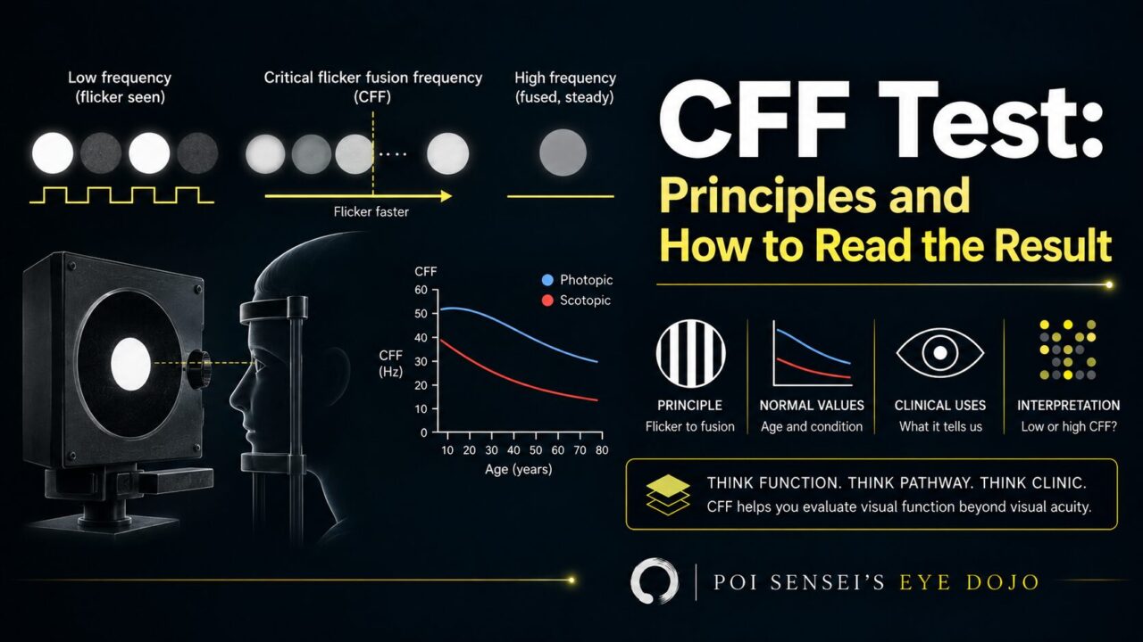
The critical flicker fusion (CFF) test is mainly used to look for problems in the optic nerve. It is quick and non-invasive.
The patient looks into the device with one eye and watches a flickering light.
- The flicker gets faster until the patient can no longer see it flicker.
- Then it starts too fast to see and slows down until the flicker appears again.
- The result is given in hertz (Hz). 35 Hz or above is normal.
Why fast flicker stops being visible
Why can’t we see a light flickering faster than about 50 Hz?
The classic answer is the refractory period: after a nerve fires, there is a short time when it cannot fire again. When flashes come faster than the nerve can answer, they blend into a steady light.
Some papers describe the limit in the healthy eye more broadly, as the temporal resolution of the visual system[1]. Where the refractory period clearly matters is the damaged nerve: in demyelinated axons it becomes 2 to 5 times longer than normal[2].
Conditions that lower the CFF value
CFF drops in optic nerve disease, such as optic neuritis and optic neuropathy[3].
In optic neuritis, the myelin is damaged, so the signal can no longer jump quickly from node to node. Conduction slows, the refractory period gets longer[2], and CFF falls.
The retina can lower CFF too, for example in cone dystrophy. Fast flicker is seen by cones, not rods. That is also why the flicker ERG tests cone function.
- Compare the two eyes. A difference of 3 Hz or more is a finding[4].
- Check the lower eye against 35 Hz, or your clinic’s own range.
- Ask where the problem is: the cones or the optic nerve.
- Ask what else could have lowered it.
Reading the number
Start with the difference between the eyes. The other eye is the best control you have. In pituitary tumors, a difference of 3 Hz or more was the best cut-off for detecting tumor contact with the optic nerve or chiasm[4].
CFF tells you that something is wrong, not why. Optic neuritis and ischemic optic neuropathy gave similar values overall (20.7 Hz and 24.3 Hz, not significantly different)[5]. CFF alone cannot reliably tell them apart.
What lowers CFF that is not the nerve
Poor fixation or unreliable answers also give a low value that means nothing. In my experience, the result is also unreliable when best-corrected acuity is 20/400 (1.3 logMAR) or worse, so read it with caution in those eyes.
Do not blame the cataract
Following a patient over time
A note for readers outside Japan
In Japan, CFF meters are standard clinic equipment, and the test is routine when optic neuropathy is suspected. In the United States, a major insurer classifies it as experimental[9]. Where there is no CFF meter, the visual evoked potential, the relative afferent pupillary defect, and color vision testing help answer a similar question.
Summary
- CFF checks the optic nerve and the cones. 35 Hz or above is normal.
- Compare the two eyes first. A 3 Hz difference matters.
- A low CFF mainly suggests optic nerve disease; cone disease can also lower it.
- A cataract rarely explains a low CFF, but take care with a mature or very dense one.
References
[1] Xu G, Fu J, Qi H, et al. The theory of critical flicker fusion frequency and its application in cataracts. Advances in Ophthalmology Practice and Research. 2023;3(1):29–32. PMID 37846427.
[2] Felts PA, Baker TA, Smith KJ. Conduction in segmentally demyelinated mammalian central axons. The Journal of Neuroscience. 1997;17(19):7267–7277. PMID 9295373.
[3] Fu J, Wang Y, Tan S, et al. The clinical application of critical flicker fusion frequency in demyelinating optic neuritis. Advances in Ophthalmology Practice and Research. 2021;1(2):100011. PMID 37846319.
[4] Taguchi A, Kinoshita Y, Tokumo K, et al. Usefulness of critical flicker fusion frequency measurement and its laterality for evaluating compressive optic neuropathy due to pituitary neuroendocrine tumors. Neurosurgical Review. 2022;46(1):4. PMID 36471083.
[5] Young MT, Braich PS, Haines SR. Critical flicker fusion frequency in demyelinating and ischemic optic neuropathies. International Ophthalmology. 2018;38(3):1069–1077. PMID 28527029.
[6] Muth T, Schipke JD, Brebeck AK, Dreyer S. Assessing critical flicker fusion frequency: which confounders? A narrative review. Medicina (Kaunas). 2023;59(4):800. PMID 37109758.
[7] Shankar H, Pesudovs K. Critical flicker fusion test of potential vision. Journal of Cataract and Refractive Surgery. 2007;33(2):232–239. PMID 17276263.
[8] Tsumura R, Harada Y, Chuman H, Kiuchi Y. Assessing the correlation between visual acuity and critical fusion frequency in acute optic neuritis before and after steroid therapy. Cureus. 2023;15(12):e49965. PMID 38179351.
[9] Aetna. Clinical Policy Bulletin 0860: Critical flicker fusion. Accessed September 3, 2026.