Poi Sensei's Eye Dojo
  • Home
  • About Poi Sensei
  • Article List
  • Contact Us
  • Japanese Site
Intorsion vs extorsion: which muscles roll the eye, and how to see torsion. A line drawing of an eyeball rolling around its visual axis, with icons for the 12 o'clock mark, the fundus and the double Maddox rod
Neuro-ophthalmology

Intorsion vs Extorsion: Which Muscles Roll the Eye, and How to See Torsion in Your Patient

September 25, 2026 JARVIS
Bielschowsky head tilt test: the eyes counter-roll when the head tilts, and in superior oblique palsy the affected eye goes up
Neuro-ophthalmology

Bielschowsky Head Tilt Test: Why the Eye Goes Up, and How to Read It

September 17, 2026 Poi Sensei
Hess chart interpretation: which muscle is weak, and is it really a palsy? Two Hess charts side by side with a smaller paretic field, a yoke muscle overacting, and an eye that will not abduct
Ophthalmic Examinations

Hess Chart Interpretation: Which Muscle Is Weak, and Is It Really a Palsy?

September 16, 2026 Poi Sensei
White dot syndromes: telling them apart on fluorescein angiography, with a fundus showing white dots and a four-step checklist — one eye or both, early FA, late FA, and not all white dots are white dot syndromes
Uveitis

White Dot Syndromes: Telling Them Apart on Fluorescein Angiography

September 15, 2026 Poi Sensei
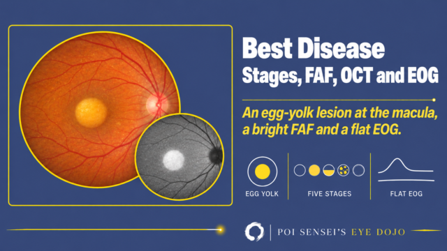
EOG test: the Arden ratio and what it tells you. Nasal and temporal electrodes, the RPE on OCT, the dark trough and light peak curve, and Best disease with an abnormal EOG and a normal ERG
Ophthalmic Examinations

EOG Test: The Arden Ratio and What It Tells You

September 7, 2026 Poi Sensei
VEP test: how to read a visual evoked potential. A checkerboard stimulus, the visual pathway and the N75-P100-N145 waveform
Ophthalmic Examinations

VEP Test: How to Read a Visual Evoked Potential

September 6, 2026 Poi Sensei
Drusen subtypes: how to tell them apart on OCT and FA. Four fundus images with matching OCT sections, and icons for position, OCT, FA and pattern
Macular Diseases

Drusen Subtypes: How to Tell Them Apart on OCT and FA

September 2, 2026 Poi Sensei
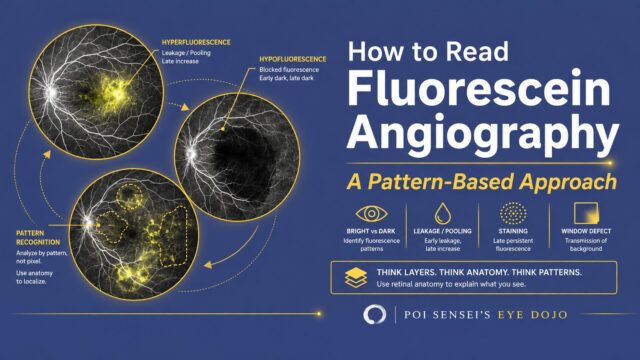
Fluorescein angiography interpretation: hyperfluorescence from leakage and pooling, hypofluorescence from blocked fluorescence, staining, and window defect
Ophthalmic Examinations

How to Read Fluorescein Angiography: A Pattern-Based Approach

August 28, 2026 Poi Sensei
Eyelid anatomy: a clinically oriented guide. A line drawing of a sagittal section of the upper eyelid, with the orbital septum highlighted
Anatomy & Physiology

Eyelid Anatomy: A Clinically Oriented Guide

May 3, 2026 Poi Sensei
Anatomy of the orbit: bones, fissures and what passes through. A line drawing of the right bony orbit from the front, with the superior orbital fissure highlighted
Anatomy & Physiology

Anatomy of the Orbit

July 21, 2025 Poi Sensei
  • 1
  • 2
Ophthalmologist & Educator
Poi Sensei
Hi, I'm Poi Sensei, a board-certified ophthalmologist in Japan. More than 2,600 Japanese eye doctors study with my content. Here I explain how to read an ERG, a fluorescein angiogram, or a visual field, in plain English. Let's learn together!
\ Follow me /
Categories
  • Anatomy & Physiology 5
  • Ophthalmic Examinations 6
  • Neuro-ophthalmology 3
  • Uveitis 1
  • Macular Diseases 1
Privacy Policy Disclaimer 2025–2026  Poi Sensei