Poi Sensei's Eye Dojo
  • Home
  • Articles
    • All articles
    • Anatomy & Physiology
    • Ophthalmic Examinations
    • Neuro-ophthalmology
    • Macular Diseases
    • Uveitis
    • Corneal and Conjunctival Diseases
    • Retinal Diseases
    • Congenital Diseases
  • About Poi Sensei
  • Contact Us
  • Japanese Site
― CATEGORY ―

Macular Diseases

Poi Sensei  October 28, 2026
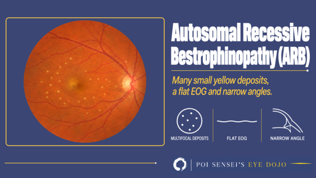
Autosomal recessive bestrophinopathy: fundus photograph with many small yellow deposits over the posterior pole
Macular Diseases

Autosomal Recessive Bestrophinopathy (ARB): Fundus, OCT and EOG

October 28, 2026 Poi Sensei
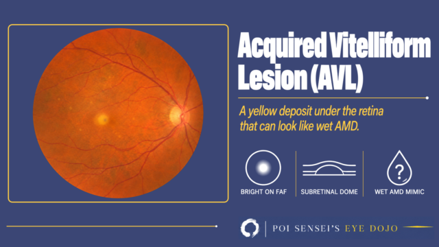
Acquired vitelliform lesion: fundus photograph with a small yellow deposit at the fovea
Macular Diseases

Acquired Vitelliform Lesion (AVL): OCT, Autofluorescence and How Not to Mistake It for Wet AMD

October 28, 2026 Poi Sensei
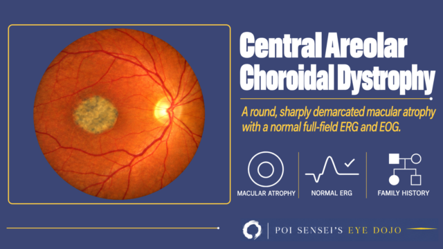
Central Areolar Choroidal Dystrophy: a fundus photograph with a round, sharply demarcated patch of macular atrophy
Macular Diseases

Central Areolar Choroidal Dystrophy (CACD): Fundus, Autofluorescence and ERG

October 28, 2026 Poi Sensei
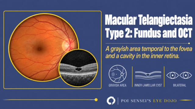
Macular telangiectasia type 2: fundus photograph with a grayish area temporal to the fovea and an OCT with an inner retinal cavity
Macular Diseases

Macular Telangiectasia Type 2 (MacTel): Fundus and OCT Findings

October 25, 2026 Poi Sensei
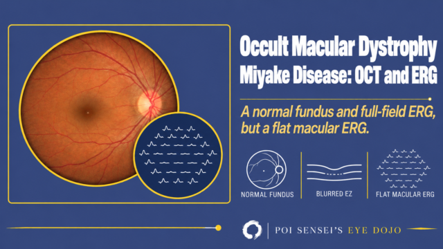
Occult macular dystrophy: a normal fundus with a multifocal ERG that is flat in the center
Macular Diseases

Occult Macular Dystrophy (Miyake Disease): OCT and ERG Findings

October 25, 2026 Poi Sensei
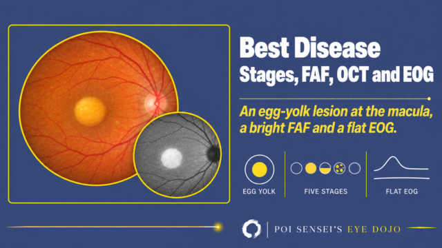
Best disease thumbnail: color fundus photograph with a round yellow egg-yolk lesion at the macula and an autofluorescence image in which the lesion is bright
Macular Diseases

Best Disease: The Five Stages on Fundus, FAF, OCT and EOG

October 23, 2026 Poi Sensei
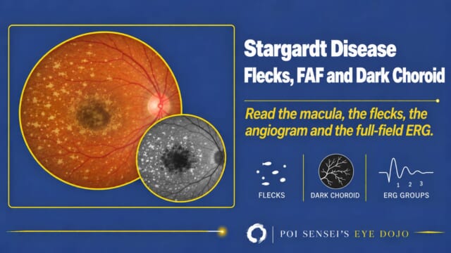
Stargardt Disease: Flecks, FAF and Dark Choroid. Illustrated fundus with macular atrophy and yellow flecks, and an autofluorescence inset
Macular Diseases

Stargardt Disease: Flecks, Autofluorescence, the Dark Choroid and ERG Groups

October 14, 2026 Poi Sensei
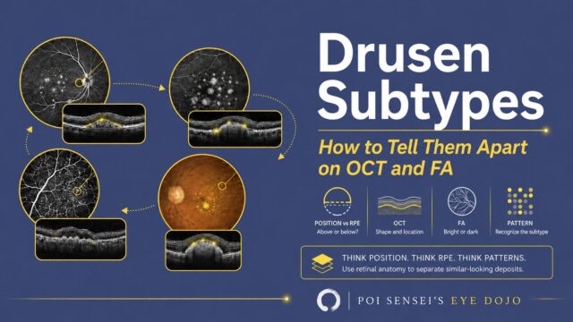
Drusen subtypes: how to tell them apart on OCT and FA. Four fundus images with matching OCT sections, and icons for position, OCT, FA and pattern
Macular Diseases

Drusen Subtypes: How to Tell Them Apart on OCT and FA

September 2, 2026 Poi Sensei
Ophthalmologist & Educator
Poi Sensei
Hi, I'm Poi Sensei, a board-certified ophthalmologist in Japan. More than 2,600 Japanese eye doctors study with my content. Here I explain how to read an ERG, a fluorescein angiogram, or a visual field, in plain English. Let's learn together!
\ Follow me /
My Book
Cover of Ophthalmology for Your First Year, Volume 1 by Poi Sensei

Ophthalmology for Your First Year, Volume 1

A plain-English guide for your first year of ophthalmology training. Kindle and paperback.

See it on Amazon
Categories
  • Anatomy & Physiology 5
  • Ophthalmic Examinations 8
  • Corneal and Conjunctival Diseases 2
  • Neuro-ophthalmology 6
  • Uveitis 2
  • Retinal Diseases 16
  • Macular Diseases 8
  • Congenital Diseases 3
  • HOME
  • Macular Diseases
Privacy Policy Disclaimer 2025–2026  Poi Sensei