
Understanding the anatomy of the lacrimal drainage system is essential for ophthalmologists, both in clinical practice and for ophthalmology exams. This article outlines the key anatomical components, measurements, and clinical relevance of each structure.
Anatomy of the Lacrimal Drainage System
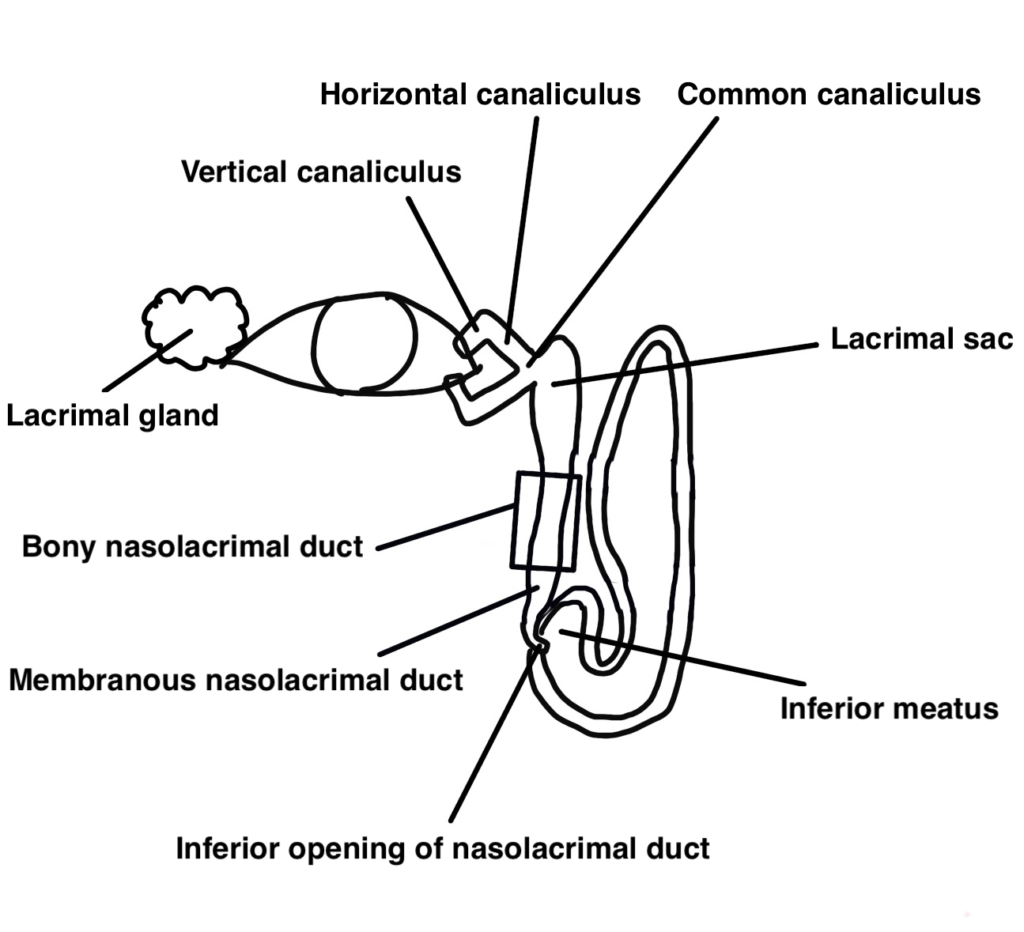
Punctum (Lacrimal Punctum)
The lacrimal puncta are located approximately 6 to 6.5 mm from the medial canthus on both the upper and lower eyelids.
A classic sign of canaliculitis is punctal dilation with redness and localized swelling.
Canaliculi (Lacrimal Canaliculi)
From the punctum, the canaliculus has a vertical segment followed by a horizontal segment, eventually merging into the common canaliculus.
- Inner diameter: 0.5 to 0.7 mm
- Epithelium: Stratified squamous epithelium
- Vertical segment: 2 mm in length
- Horizontal segment: 10 mm in length
- Common canaliculus: Inner diameter 0.9 to 1.2 mm, which opens into the lacrimal sac
Lacrimal Sac
- Length: 10 mm
- Depth: 7–8 mm
- Width: 1–2 mm
- Medial wall (posterior): Adjacent to bone
- Lateral wall (anterior): Covered by orbital septum, orbicularis oculi, and medial canthal ligament
- Epithelium: Lined with pseudostratified columnar epithelium and basal cells
The distance from the punctum to the nasal wall of the lacrimal sac is about 12–16 mm (average 14 mm)
Nasolacrimal Duct
The nasolacrimal duct continues from the lacrimal sac through the membranous nasolacrimal duct and opens into the inferior meatus of the nasal cavity.
Importantly:
- The upper membranous portion is enclosed by the bony nasolacrimal canal, which consists of the lacrimal bone and maxilla.
- Some literature incorrectly describes the lacrimal sac as connecting directly to the bony duct and then to the membranous part—this is anatomically inaccurate and should be noted.
- Total length of the nasolacrimal duct: 17 mm
- The full distance from the punctum to the lower nasal opening ranges from 30 to 45 mm, varying among individuals.
- Around 90% of tears are reabsorbed through the nasolacrimal duct, while only 10% drain into the nasal cavity.
Clinical Relevance
Precise anatomical knowledge of the lacrimal drainage system is frequently tested in ophthalmology exams, especially the lengths and spatial relationships between components.
In clinical settings, such knowledge is also vital—for example, during bougie probing or lacrimal surgery, knowing how far to advance the instrument to reach the lacrimal sac is crucial.
One useful tip is to visualize these distances on your own face to build spatial awareness. This approach not only helps with memory retention but also improves practical skills during procedures.
Summary
The lacrimal drainage system is a small but intricate anatomical pathway, with several clinically significant variations. A solid grasp of its structure is essential for both examination success and effective patient care in lacrimal disorders.