Bielschowsky Head Tilt Test: Why the Eye Goes Up, and How to Read It
What you will be able to do
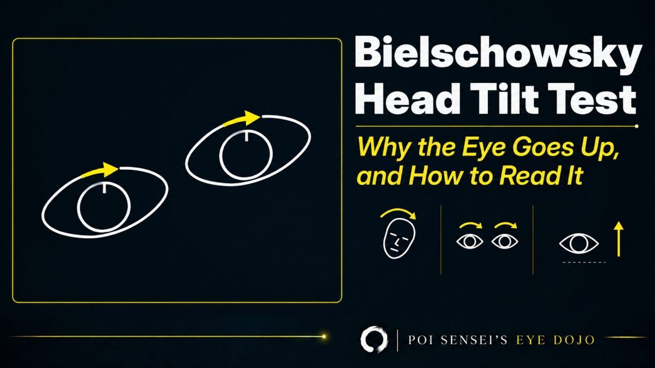
Every textbook says the same thing about the Bielschowsky head tilt test: in a superior oblique palsy, tilt the head toward the affected side and the affected eye goes up. Most people memorize that sentence and stop. This article shows you why it happens, so you can derive it from the anatomy instead, and shows the second diagnosis the same finding can point to. It ends with the two situations where the test lets you down.
What the eyes do when the head tilts
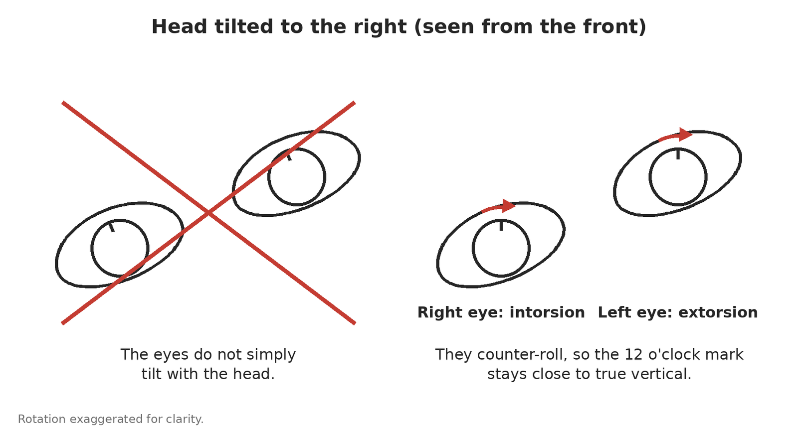
Tilt the head to the right and the eyes do not simply tilt with it. They counter-roll: the right eye intorts and the left eye extorts, so that the 12 o’clock point on each cornea stays closer to true vertical. This counter-roll is the whole basis of the test.
Now recall which muscles do the rolling. Intorsion: superior oblique and superior rectus. Extorsion: inferior oblique and inferior rectus. If those pairs are not automatic yet, our guide to extraocular muscle actions covers them. Each pair is one oblique plus one rectus, and the rectus in each pair also moves the eye vertically. That detail is what makes the test work.
Why the affected eye goes up
Take a right superior oblique palsy and tilt the head to the right. Follow one eye at a time.
- The left eye extorts as usual. Nothing is wrong on that side.
- The right eye has to intort. It has two intorters: the superior oblique and the superior rectus.
- The superior oblique is paretic. So the superior rectus has to do the intorting alone.
- But the superior rectus is also an elevator. When it contracts, it intorts and elevates at the same time.
- So the right eye goes up.
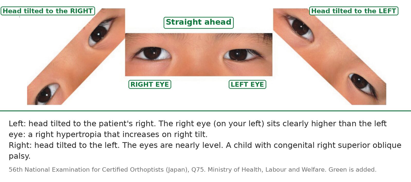
The Bielschowsky head tilt test is positive: a right hypertropia on right head tilt.
Tilt the head to the left instead and the right eye has to extort, which the inferior oblique and inferior rectus can still do. Nothing is asked of the paretic muscle, so the hypertropia shrinks.
This is what it looks like in a child with congenital right superior oblique palsy:
The mirror image: same finding, different muscle
Here is the question worth asking: does the same logic work for extorsion?
It does. Suppose the left inferior oblique is paretic. This is rare, because the inferior oblique shares the third nerve with several other muscles, so it is not widely taught. Tilt the head to the right and follow the left eye.
- The right eye intorts as usual. Nothing is wrong on that side.
- The left eye has to extort. It has two extorters: the inferior oblique and the inferior rectus.
- The inferior oblique is paretic. So the inferior rectus has to do the extorting alone.
- But the inferior rectus is also a depressor. When it contracts, it extorts and depresses at the same time.
- So the left eye goes down.
A left eye that goes down is, from the examiner’s side, the same picture as a right eye that goes up: a right hypertropia that increases on right head tilt. So a positive tilt to the right does not by itself mean right superior oblique palsy. It means right superior oblique palsy or left inferior oblique palsy. The tilt alone cannot tell them apart, which is exactly why Parks combined it with two other steps[1].
Learn the test from the mechanism and you will not forget it. Learn it as a sentence and you will.
Where it sits in the three-step test
The head tilt is the third step of the Parks three-step test[1]:
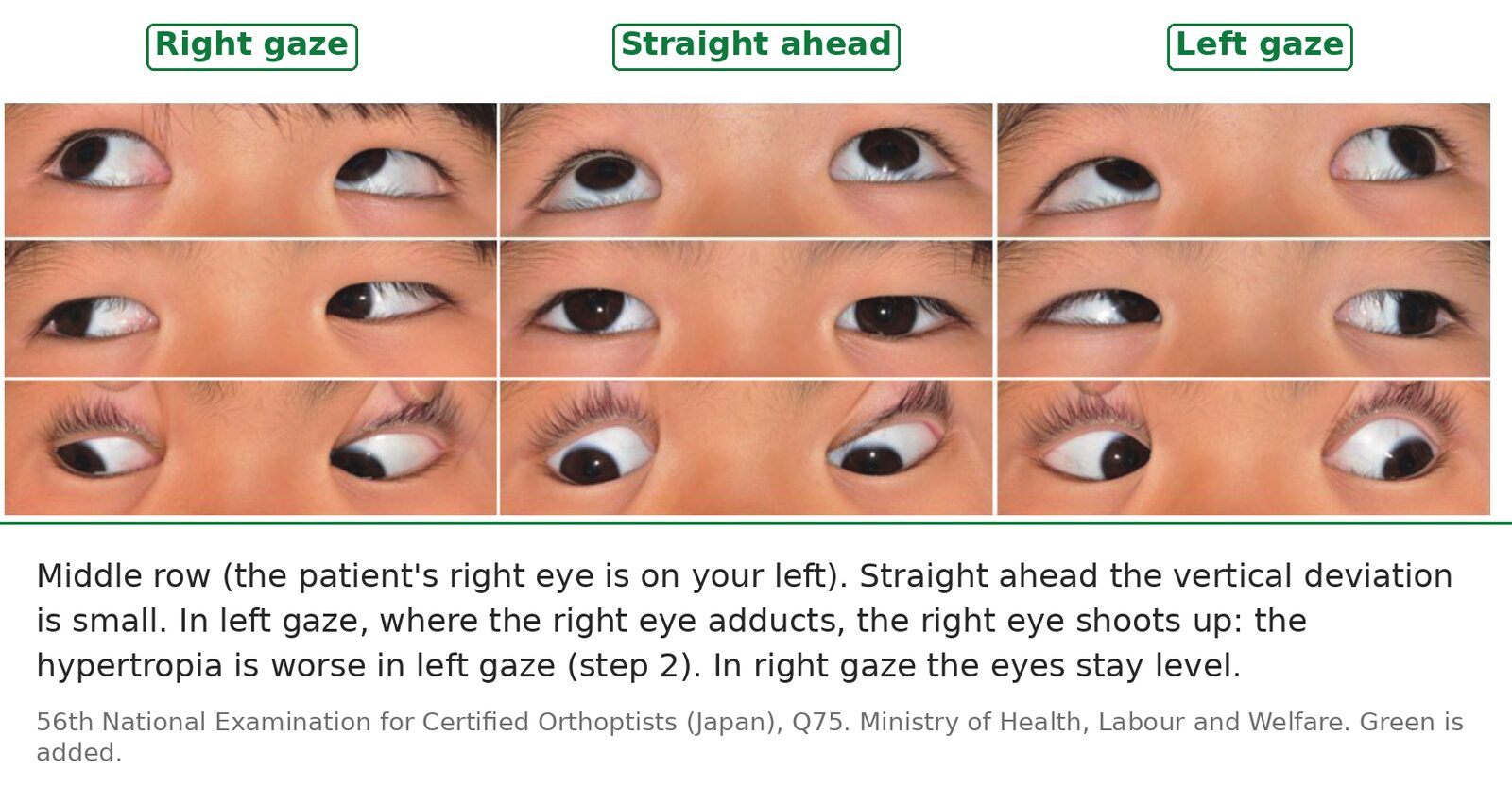
- Which eye is higher in primary position? A right hypertropia means a weak depressor of the right eye (superior oblique, inferior rectus) or a weak elevator of the left eye (superior rectus, inferior oblique). Four muscles.
- Is the hypertropia worse in right gaze or left gaze? Two muscles.
- Worse on right tilt or left tilt? One muscle.
Each step halves the list, and the tilt is the step that separates an oblique from a rectus. The same child shows steps 1 and 2:
Two situations where it lets you down
Bilateral palsy after head trauma. Superior oblique palsy after head trauma is often bilateral, and this is where the head tilt test is weakest: in one series of proven bilateral cases it was positive in fewer than half[3]. If the history is trauma, look for the bilateral signs instead: a V pattern, extorsion that is greater in downgaze than in upgaze, and extorsion of both fundi[3].
Sagging eye syndrome in older patients. An older patient with a small vertical deviation and diplopia may have no nerve palsy at all. In sagging eye syndrome the deviation barely changes between the two tilts, about 2 prism diopters against about 14 in trochlear nerve palsy, and a change of 6 prism diopters or more argues against sagging eye syndrome[4]. A small deviation that hardly changes with tilt, in an older patient, is not a fourth nerve palsy.
Summary
- Head tilt makes the eyes counter-roll: the same-side eye intorts, the opposite eye extorts.
- Intorters: superior oblique + superior rectus. Extorters: inferior oblique + inferior rectus. The rectus in each pair also moves the eye vertically.
- In right superior oblique palsy, right tilt forces the superior rectus to intort alone, and it elevates: the right hypertropia increases.
- The same finding also fits a left inferior oblique palsy. The tilt alone cannot separate them[1].
- The test misses about 3 in 10 unilateral palsies[2] and most bilateral ones[3]. In older patients, a small deviation that hardly changes with tilt points to sagging eye syndrome[4].
References
[1] Parks MM. Isolated cyclovertical muscle palsy. AMA Archives of Ophthalmology. 1958;60(6):1027–1035. PMID 13593934.
[2] Manchandia AM, Demer JL. Sensitivity of the three-step test in diagnosis of superior oblique palsy. Journal of AAPOS. 2014;18(6):567–571. PMID 25459202.
[3] Muthusamy B, Irsch K, Peggy Chang HY, Guyton DL. The sensitivity of the Bielschowsky head-tilt test in diagnosing acquired bilateral superior oblique paresis. American Journal of Ophthalmology. 2014;157(4):901–907.e2. PMID 24412122.
[4] Yamadera K, Kimura A, Okita Y, Mochizuki Y, Gomi F. Comparison of head tilt test between sagging eye syndrome and acquired unilateral trochlear nerve palsy. Graefe’s Archive for Clinical and Experimental Ophthalmology. 2024;262(5):1641–1646. PMID 38141057.
Figure credits
Photographs reproduced from the 56th National Examination for Certified Orthoptists (Japan), question 75, published by the Ministry of Health, Labour and Welfare. Anything in green is mine. The counter-roll diagram is mine.