Intorsion vs Extorsion: Which Muscles Roll the Eye, and How to See Torsion in Your Patient

What you will be able to do
Many people find torsion the hardest part of eye movements. Intorsion and extorsion are easy to define, yet easy to mix up when a patient is in front of you. This article gives you one rule for the direction, one rule for the muscles, and then the part most textbooks skip: how to actually see torsion in your patient, on a fundus photograph and with a double Maddox rod, and what it means when the two disagree.
Step 1: Which way is which
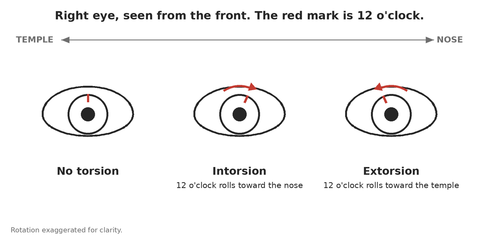
Put a mark at 12 o’clock on the cornea and watch where it goes.
- Intorsion: the 12 o’clock mark rolls toward the nose.
- Extorsion: the 12 o’clock mark rolls toward the temple.
That is the whole definition. The direction is always named from the top of the eye. Torsion of one eye is also called incyclotorsion and excyclotorsion, and a torsional deviation is an incyclotropia or excyclotropia. Same thing.
Step 2: Which muscles roll the eye
The obliques do most of the rolling. The superior oblique intorts and the inferior oblique extorts. The vertical recti add a little: the superior rectus intorts and the inferior rectus extorts. So:
- Intorters: superior oblique and superior rectus.
- Extorters: inferior oblique and inferior rectus.
Notice the pattern. The two muscles that attach to the top of the globe intort. The two that attach to the bottom extort. The reason is that all four pull their insertion toward the nose: the recti pull back toward the orbital apex, which sits nasal to the eye’s axis, and the obliques pull toward the trochlea and the nasal orbital floor. Pull the top of the eye nasally and 12 o’clock rolls toward the nose. Pull the bottom nasally and 12 o’clock rolls toward the temple.
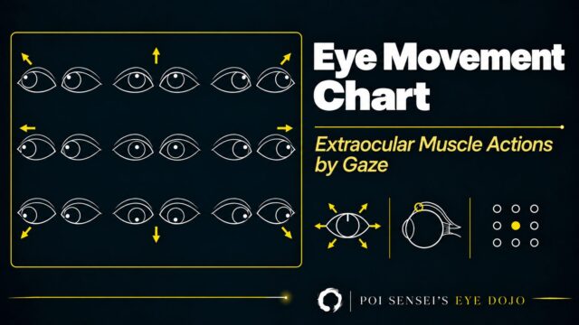
How much each muscle rolls the eye, and how much it moves it up or down, changes with the direction of gaze. That is a separate topic and we cover it in our guide to extraocular muscle actions.
Step 3: Which way a palsied eye rolls, and what the patient sees
Lose an intorter and the eye extorts. A superior oblique palsy therefore gives an eye that sits extorted and, because the superior oblique is also a depressor, higher than the other eye.
Now the part people get backwards. The eye rolls one way; the world rolls the other way. If the right eye is extorted, the image the right eye sees appears intorted, tilted toward the nose. The patient does not report "my eye is rolled." They report that horizontal things are no longer horizontal.
If this is hard to picture, try it with your phone. Hold it in front of your right eye and let the camera be the eye.
- Take a photo looking straight at something horizontal, such as a shelf or a door frame.
- Now roll the phone into extorsion. For a right eye, that is clockwise as you see it. Take the photo again.
- Compare the two. In the second photo the shelf is tilted counterclockwise: the eye rolled out, so the image rolled in.
The same trick works for any deviation. Turn the phone outward, like an exotropic right eye, and the object moves to the left of the photo. That is crossed diplopia.
Now a real patient.
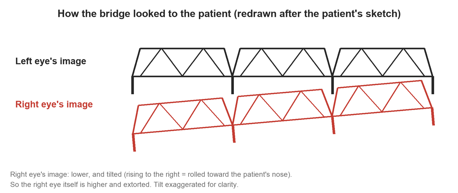
A 65-year-old man with diabetes and no history of trauma noticed, a week ago, that a bridge over the river looked tilted. He brought a drawing of how it looked.
- In his drawing, the right eye’s image sits lower and is tilted, rising to the right.
- The right eye’s image is intorted, so the right eye itself is extorted. The image sits lower, so the right eye is higher.
- That is a right superior oblique palsy. With diabetes and no trauma, the cause is most likely ischemic.
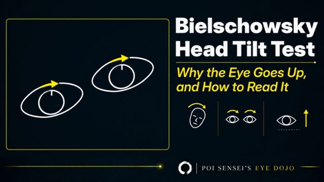
Tilting the head to the left, away from the palsy, relieves the symptom; why that works is the Bielschowsky head tilt test.
Step 4: How to see torsion
There are two kinds of torsion, and you need both.
Objective torsion is what the eye actually did. You see it on the fundus. The fovea normally sits a little below the center of the optic disc: on fixation photographs of 50 non-strabismic subjects, 0.3 disc diameters below a horizontal line through the disc center[1]. As an angle between that horizontal line and the disc-to-fovea line, it is about 7° in normal children, and the measurement is highly reproducible between observers[2].
- Extorsion: the fovea drops further below the disc center. The angle grows.
- Intorsion: the fovea rises to the level of the disc center or above it. The angle shrinks or reverses.
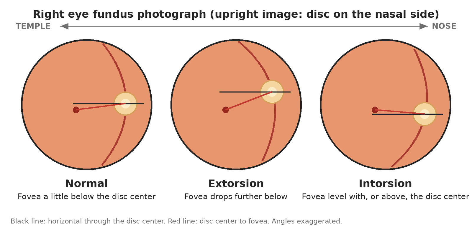
You can see the same thing at the slit lamp with a fundus lens, but a photograph lets you measure it and compare visits. Photograph both eyes[3].
Subjective torsion is what the patient perceives. The double Maddox rod test measures it. A red Maddox rod goes in front of one eye and a white one in front of the other, both with the cylinders vertical, so the patient sees two horizontal lines. If one line is tilted, the patient rotates that rod until the lines are parallel, and the degrees on the trial frame are the subjective torsion.
Let’s look at a real case.
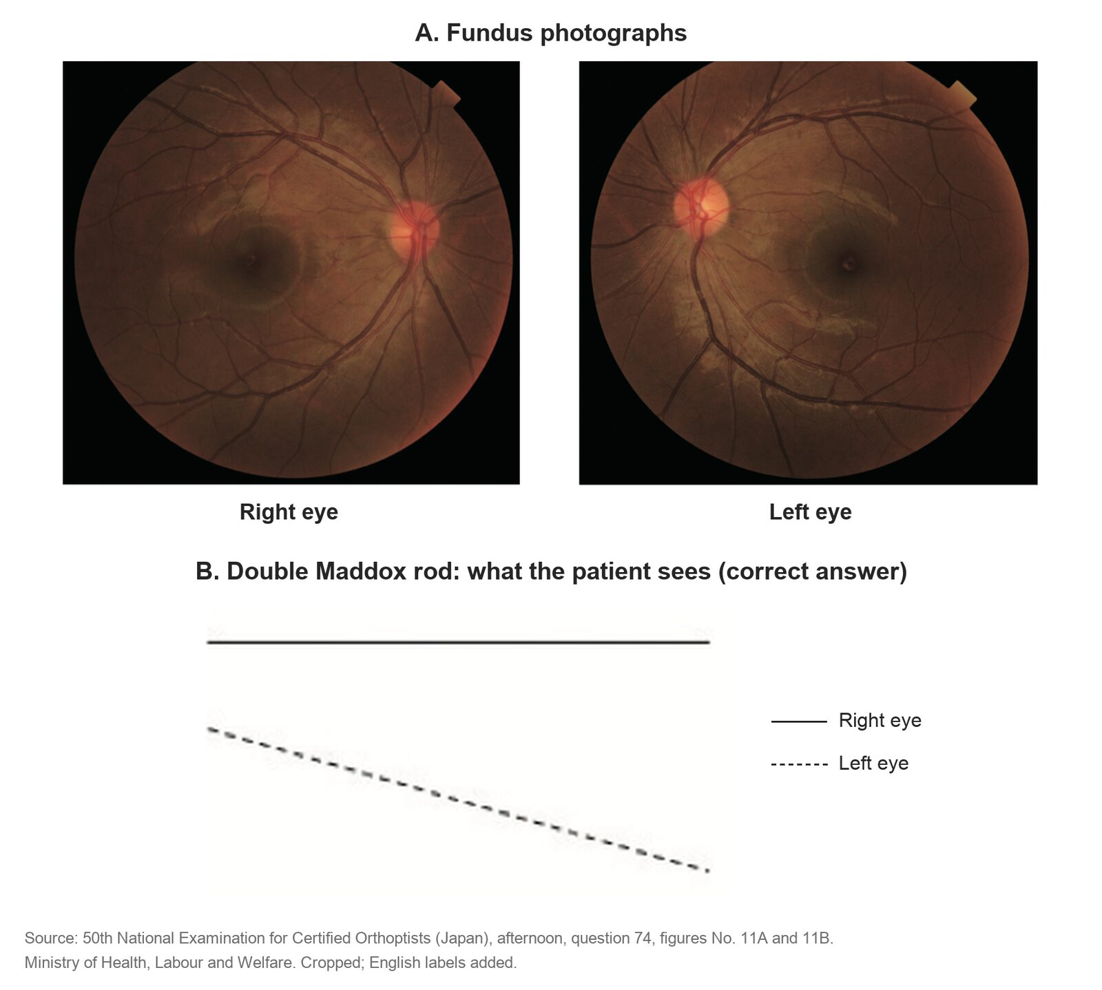
A 38-year-old man hit his head in a traffic accident, lost consciousness briefly, and then noticed double vision. He has a left hypertropia.
- Fundus photographs: in the left eye, the fovea sits well below the disc center. The left eye is extorted.
- Double Maddox rod: the right eye’s line is horizontal. The left eye’s line is lower and tilted, sloping down to the patient’s right.
- The left eye rolls out, so the line it sees rolls in. That is a left superior oblique palsy after head trauma.
When the fundus and the patient disagree
Summary
There are only three things to remember about torsion.
- Name the direction by 12 o’clock. Toward the nose is intorsion; toward the temple is extorsion.
- Top muscles intort, bottom muscles extort, because all four pull their insertion toward the nose.
- When the eye rolls one way, the image rolls the other way. A right eye that sees the world intorted is itself extorted.
In clinic, if you suspect torsion, photograph the fundus of both eyes. Even when the patient does not notice it, the fundus shows it.
References
[1] Bixenman WW, von Noorden GK. Apparent foveal displacement in normal subjects and in cyclotropia. Ophthalmology. 1982;89(1):58–62. PMID 7070775.
[2] Le Jeune C, Chebli F, Leon L, Anthoine E, Weber M, Péchereau A, Lebranchu P. Reliability and reproducibility of disc-foveal angle measurements by non-mydriatic fundus photography. PLoS One. 2018;13(1):e0191007. PMID 29370195.
[3] Kim DH, Lim HT. Comparison of ocular torsion between congenital and acquired unilateral superior oblique palsy. Eye (London). 2019;33(10):1658–1663. PMID 31171838.
[4] Kawai M, Goseki T, Okano T, Ishikawa H. Comparison of subjective cyclofusion ranges and objective ocular torsion in normal participants according to age. Graefe’s Archive for Clinical and Experimental Ophthalmology. 2022;260(11):3675–3681. PMID 35708848.
[5] Liebermann L, Hatt SR, Leske DA, Klaehn LD, Kramer AM, Holmes JM. Comparison of methods for measuring cyclodeviation. American Journal of Ophthalmology. 2021;224:332–342. PMID 33253661.
[6] Roh YR, Hwang JM. Comparison of subjective and objective torsion in patients with acquired unilateral superior oblique muscle palsy. British Journal of Ophthalmology. 2011;95(11):1583–1587. PMID 21427460.
[7] Muthusamy B, Irsch K, Peggy Chang HY, Guyton DL. The sensitivity of the Bielschowsky head-tilt test in diagnosing acquired bilateral superior oblique paresis. American Journal of Ophthalmology. 2014;157(4):901–907.e2. PMID 24412122.