Achromatopsia: Symptoms, Fundus, OCT and ERG Findings
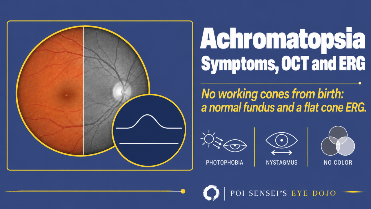
A baby has poor vision, nystagmus and strong discomfort in bright light, but the fundus looks normal. Think of achromatopsia. This article explains what achromatopsia is, and how to read the OCT and ERG that make the diagnosis.
- Poor vision, nystagmus and photophobia are present from infancy.
- The patient sees no colors at all.
- The fundus looks normal.
- On the ERG, the cone response and 30 Hz flicker are absent, while the rod response is normal.
What is achromatopsia?
Achromatopsia is a congenital disease in which all three types of cones (red, green and blue) do not work from birth. The rods work normally. In a milder form, incomplete achromatopsia, some cone function and color vision remain. It is also called rod monochromatism or total color blindness.
Inheritance
Achromatopsia is autosomal recessive. Autosomal recessive inheritance is common among inherited retinal diseases. It affects about 1 in 30,000 people, and two genes, CNGA3 and CNGB3, cause most cases[1].
Symptoms
The main symptoms are poor vision, photophobia (discomfort in bright light), day blindness (hemeralopia; worse vision in bright light), nystagmus and a lack of color vision. Corrected visual acuity is around 20/200 (6/60). All colors look like shades of gray[2].
Nystagmus appears in diseases where the macula does not work from birth. Cones are concentrated in the macula, so achromatopsia causes nystagmus too.
On the Ishihara test, patients can read only the first plate, a demonstration plate that people with color vision defects can also read.
Fundus
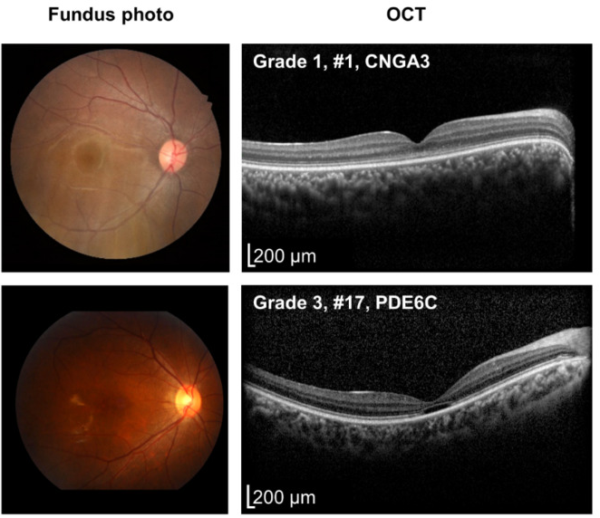
The fundus is usually normal. Some patients develop atrophy at the macula over time.
OCT
OCT can look normal, so a normal OCT does not rule out achromatopsia. OCT findings vary. The ellipsoid zone (EZ) at the fovea can be continuous, broken, or replaced by a dark gap (a hyporeflective zone). Some patients show outer retinal atrophy[1].
ERG findings
The ERG is the key test. The cone response and the 30 Hz flicker are absent or severely reduced, while the rod response is normal.
DA means dark-adapted and LA means light-adapted. The numbers 0.01 and 3.0 are the flash strength.
| ERG step | Achromatopsia |
|---|---|
| Rod (DA 0.01) | Normal |
| Mixed response (DA 3.0) | Normal or mildly reduced |
| Cone (LA 3.0) | Absent or minimal |
| 30 Hz flicker | Absent or minimal |
The 30 Hz flicker tests the cones only, because rods cannot follow 30 flashes a second.
The mixed response comes from rods and cones together, but after dark adaptation most of it comes from the rods. So it stays normal or is only mildly reduced[2].
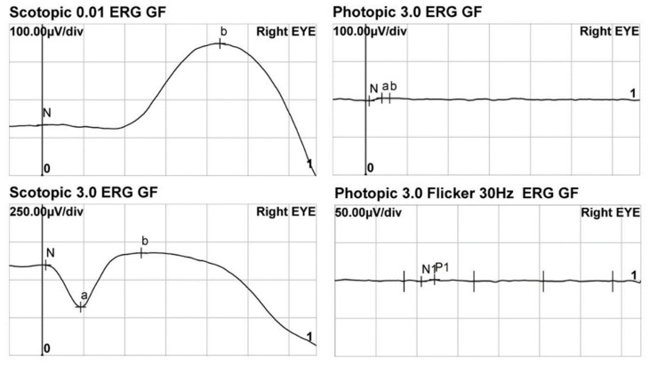
On the ERG alone, achromatopsia looks much like cone dystrophy. The history usually tells them apart. In cone dystrophy, vision is good in early childhood and falls later, usually before age 20. In achromatopsia, vision is poor from birth.
Blue cone monochromatism looks similar too, but it is X-linked, affects boys, and the blue (S) cone ERG is preserved[1].
Treatment and prognosis
There is no approved treatment. Tinted glasses help with photophobia and day blindness. Deep red tints are often used[1]. Gene therapy for CNGA3 and CNGB3 is being tested in early clinical trials. Only patients with a variant in one of these genes can join, so genetic testing is important[3].
Vision usually stays about the same, but slow cone loss over time has been reported in some patients[1].
Summary
- In achromatopsia, all three types of cones do not work from birth. The rods are normal.
- Poor vision, photophobia, day blindness, nystagmus and no color vision start in infancy.
- The fundus is usually normal, and OCT can be normal too.
- ERG: the cone response and 30 Hz flicker are absent, while the rod response is normal.
- There is no approved treatment. Tinted glasses help with photophobia.
Figure credits
Fundus, OCT and ERG: Figures 2 and 3 from Choi YJ, Joo K, Lim HT, et al. Genes 2023;14(2):519 (https://doi.org/10.3390/genes14020519), licensed under CC BY 4.0. Figure 2 cropped to two of four rows, with the row letters removed. Figure 3 cropped to four of six panels and rearranged.
References
[1] Aboshiha J, Dubis AM, Carroll J, Hardcastle AJ, Michaelides M. The cone dysfunction syndromes. Br J Ophthalmol. 2016;100(1):115–121. PMID 25770143.
[2] Salmon JF. Kanski’s Clinical Ophthalmology: A Systematic Approach. 10th ed. Elsevier; 2025. Chapter 15, Inherited retinal degenerations: Congenital achromatopsia.
[3] Baxter MF, Borchert GA. Gene therapy for achromatopsia. Int J Mol Sci. 2024;25(17):9739. PMID 39273686.
Which retinal layer makes which wave, the three abnormal patterns, and the dark-adaptation trap to check before you report “negative” — on one page, by email. One email when I publish something new, about once a month. Unsubscribe anytime.