Poi Sensei's Eye Dojo
  • Home
  • Articles
    • All articles
    • Anatomy & Physiology
    • Ophthalmic Examinations
    • Neuro-ophthalmology
    • Macular Diseases
    • Uveitis
  • About Poi Sensei
  • Contact Us
  • Japanese Site

All Articles

Poi Sensei

Every guide on Poi Sensei’s Eye Dojo, grouped by topic.

Ophthalmic Examinations 6 Anatomy & Physiology 5 Neuro-ophthalmology 4 Macular Diseases 1 Uveitis 1

Ophthalmic Examinations

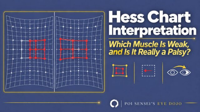
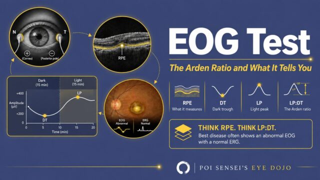
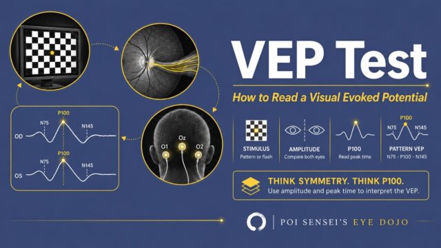
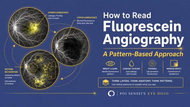
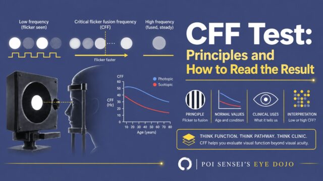
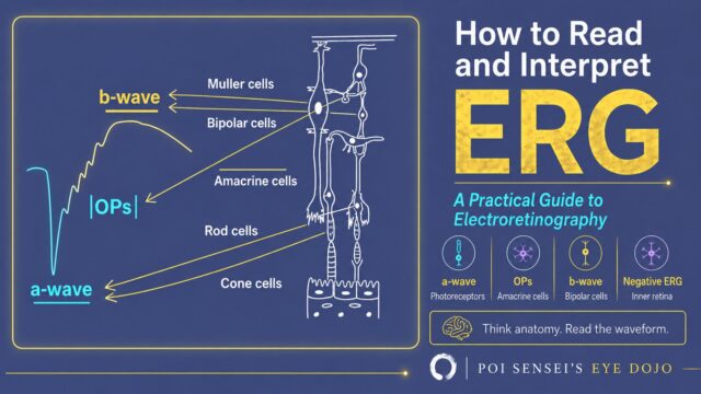
Hess chart interpretation: which muscle is weak, and is it really a palsy? Two Hess charts side by side with a smaller paretic field and an overacting yoke muscleHess Chart Interpretation EOG test: the Arden ratio and what it tells you. The eye and RPE, the dark trough and light peak on the Arden curve, and icons for RPE, DT, LP and the ratioEOG Test VEP test: how to read a visual evoked potential. A checkerboard stimulus, the visual pathway and the N75-P100-N145 waveformVEP Test How to read fluorescein angiography: a pattern-based approach. Three fluorescein angiograms showing hyperfluorescence, hypofluorescence and pattern recognitionHow to Read Fluorescein Angiography CFF test: principles and how to read the result. A flicker stimulus, the critical flicker fusion frequency and a normal-value chart by ageCFF Test How to read and interpret ERG: a practical guide to electroretinography, with the a-wave, b-wave and oscillatory potentials matched to the retinal layersERG in Ophthalmology

Anatomy & Physiology

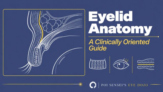
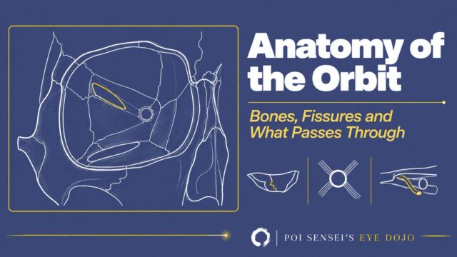
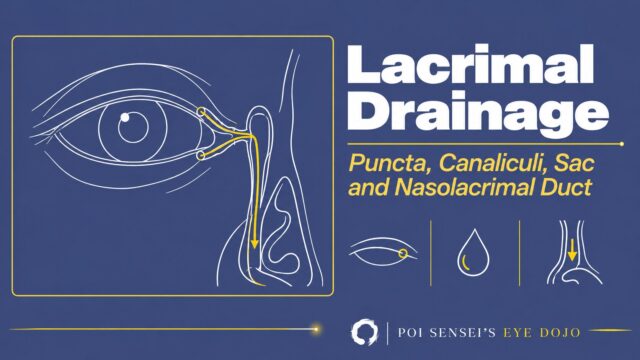
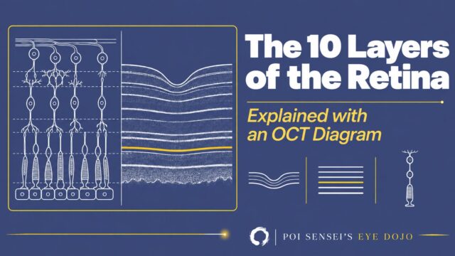
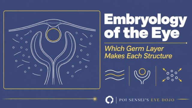
Eyelid anatomy: a clinically oriented guide. A line drawing of a sagittal section of the upper eyelid, with the orbital septum highlightedEyelid Anatomy Anatomy of the orbit: bones, fissures and what passes through. A line drawing of the right bony orbit from the front, with the superior orbital fissure highlightedAnatomy of the Orbit Lacrimal drainage system: puncta, canaliculi, lacrimal sac and nasolacrimal duct. A line drawing of the right eye with the tear drainage path highlightedAnatomy of the Lacrimal Drainage System The 10 layers of the retina, explained with an OCT diagram. Line drawings of retinal neurons beside the matching OCT bands, with the ellipsoid zone highlightedThe 10 Layers of the Retina Embryology of the eye: which germ layer makes each structure. A line drawing of the optic cup, lens vesicle and surface ectoderm, with neural crest cells around themEmbryological Origins of Eye Structures

Neuro-ophthalmology

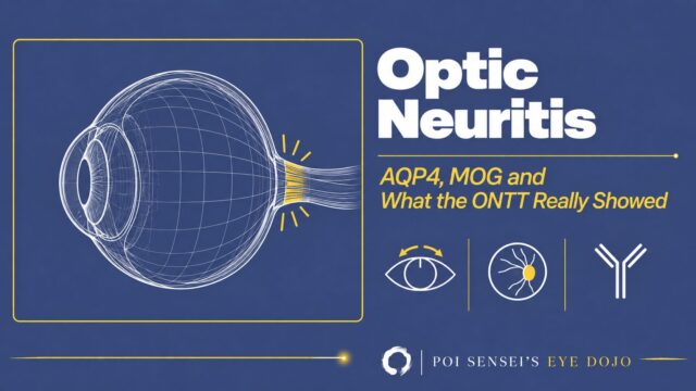
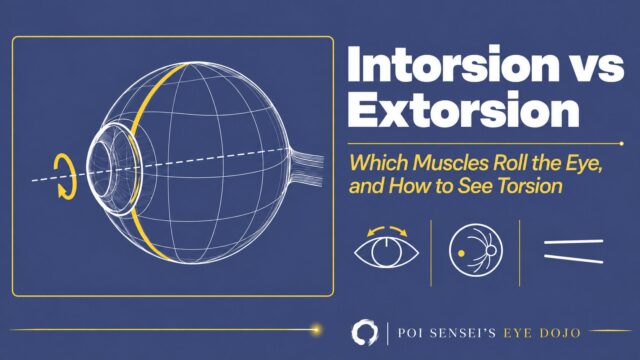
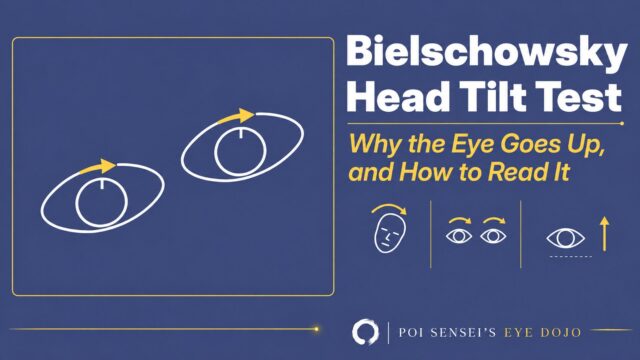
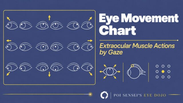
Optic neuritis: AQP4, MOG and what the ONTT really showed. A line drawing of an eyeball with the optic nerve behind it highlighted in yellow, with icons for pain on eye movement, a swollen optic disc and an antibodyOptic Neuritis Intorsion vs extorsion: which muscles roll the eye, and how to see torsion. A line drawing of an eyeball rolling around its visual axis, with icons for the 12 o'clock mark, the fundus and the double Maddox rodIntorsion vs Extorsion Bielschowsky head tilt test: the eyes counter-roll when the head tilts, and in superior oblique palsy the affected eye goes upBielschowsky Head Tilt Test Eye movement chart: extraocular muscle actions by gaze. Line drawings of both eyes in the nine positions of gaze, with arrows showing each directionEye Movement Chart

Macular Diseases

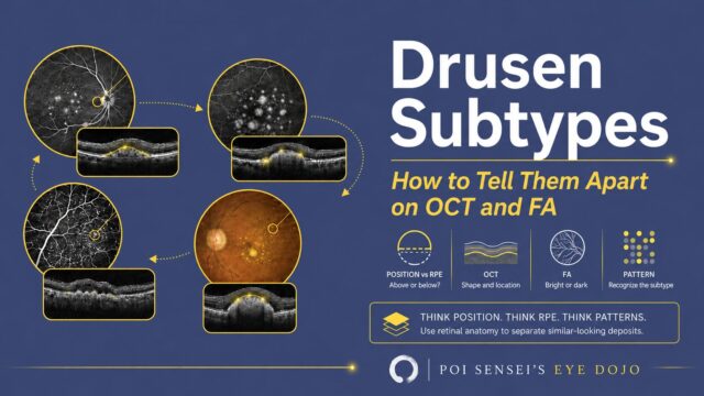
Drusen subtypes: how to tell them apart on OCT and FA. Four fundus images with matching OCT sections, and icons for position, OCT, FA and patternDrusen Subtypes

Uveitis

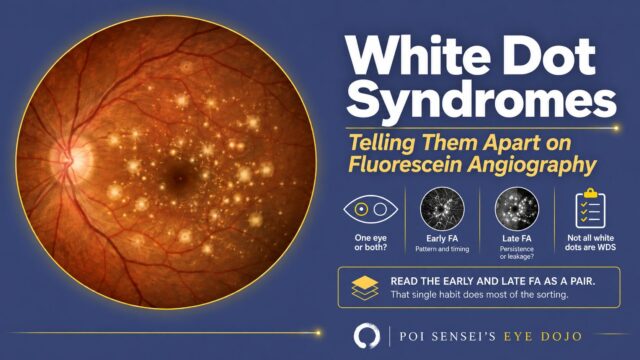
White dot syndromes: telling them apart on fluorescein angiography. A fundus photograph with scattered white dots, and icons for laterality, early FA, late FA and a checklistWhite Dot Syndromes
  • HOME
  • All Articles
Privacy Policy Disclaimer 2025–2026  Poi Sensei