White Dot Syndromes: Telling Them Apart on Fluorescein Angiography
Why they are easy to mix up
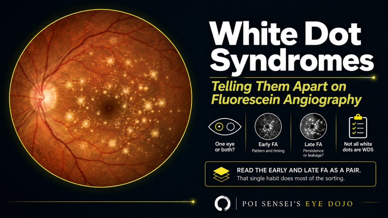
Many doctors find the white dot syndromes confusing. MEWDS, APMPPE, PIC, birdshot — there are a lot of names, and on a fundus photo most of them look alike: white or yellow-white spots in the posterior pole of a young patient.
This article does not try to cover everything. It gives you the minimum you need to tell them apart, as clearly as possible.
The key is fluorescein angiography (FA): what the lesions do in the early frames, and what they do in the late frames.
- One eye or both? That splits the group before you image anything.
- Early FA frames — are the lesions hyperfluorescent or hypofluorescent?
- Late FA frames — do they stay bright, or do they become bright?
- Ask whether this is a white dot syndrome at all. Some retinal diseases produce white dots and are not in this group.
The reversal that does most of the work
Here is the single most useful pattern.
Most of the white dot syndromes are hyperfluorescent from the early frames and remain hyperfluorescent late. MEWDS does this. PIC does this.
APMPPE does the opposite in the early frames. The lesions block early and then stain late — hypofluorescent at the start, hyperfluorescent at the end. That reversal is the finding, and it is distinctive enough that the international classification criteria adopted it: lesions that "block early and stain late diffusely" are part of the diagnostic criteria for APMPPE[1].
The reason is where the lesions sit and what they are. The APMPPE plaques are described as placoid choroidal lesions[1]; early on they mask the underlying fluorescence, and late they take up dye and stain.
The bilateral group
APMPPE — acute posterior multifocal placoid pigment epitheliopathy
Young adults, roughly 20 to 30, with bilateral acute visual loss. The anterior segment is normal.
The fundus shows scattered yellow-white patches, each about a quarter to a half disc diameter, thought to arise from edema secondary to ischemia of the RPE and outer retina. On OCT a section through a lesion shows hyper-reflectivity in the outer retina. The lesions do not enlarge or coalesce.
FA gives the reversal above. ICGA shows hypofluorescence. On autofluorescence the lesions themselves are hypoautofluorescent with a hyperautofluorescent surround.
The prognosis is good: most cases resolve spontaneously over a few weeks. Steroids are sometimes used in severe cases or to shorten the course.
PIC — punctate inner choroiditis
Young myopic women, again roughly 20 to 40. Usually bilateral, though it can present in one eye. It is not a rare disease, and it belongs on the list whenever a young woman presents with acute visual loss.
Symptoms are acute visual loss, central scotoma and photopsia.
The fundus shows yellow-white patches concentrated at the posterior pole, with little in the periphery, which scar over time. Two features separate PIC from the rest: the spots are small, and their margins are well defined. The classification criteria put a number on "small" — punctate choroidal spots under 250 µm in diameter — together with involvement of the posterior pole with or without the mid-periphery, and absent to minimal anterior chamber and vitreous inflammation[3].
That last point matters clinically: PIC is an inflammatory disease that does not show inflammation where you usually look for it.
FA shows the lesions hyperfluorescent from early to late; ICGA shows hypofluorescence.
Acute treatment is systemic steroids. The longer-term problem is that the macular lesions become atrophic scars, and type 2 choroidal neovascularization can arise from those scars — which is treated with anti-VEGF.
The rest of the bilateral group
Birdshot chorioretinopathy, multifocal choroiditis (which can also be unilateral), and serpiginous choroiditis complete the bilateral group. Each deserves its own discussion; the point here is that they belong on the list.
The unilateral group
MEWDS — multiple evanescent white dot syndrome
Myopic women, 20 to 40, with unilateral sudden visual loss and photopsia. The cause is unknown, but a preceding viral illness is common enough that a viral trigger is suspected.
The anterior segment is normal. The fundus shows white dots in the posterior pole centered around the optic nerve, roughly 100 to 200 µm, some of which coalesce, usually with disc edema. The classification criteria add a feature worth looking for specifically: foveal granularity, alongside the multifocal gray-white chorioretinal spots[2].
- FA: hyperfluorescent from early to late. The criteria describe the pattern as "wreath-like" hyperfluorescent lesions[2].
- ICGA: the spots are hypofluorescent.
- Autofluorescence: the spots are hyperautofluorescent. When the dots are hard to see on the fundus, autofluorescence shows them more clearly — worth taking for that reason alone.
- OCT: loss of the ellipsoid zone at the macula. The criteria describe hyper-reflective lesions extending from the RPE through the ellipsoid zone into the outer nuclear layer[2], which places the disease in the outer retina rather than deep in the choroid.
Inflammation in the anterior chamber and vitreous is absent to mild[2].
Treatment is usually observation. The dots disappear over about a month, the ellipsoid zone recovers, and vision usually returns. Steroid pulse therapy is sometimes given when the visual loss is severe, but its benefit is not established.
Acute retinal pigment epitheliitis
The other classically unilateral member of the group.
White dots that are not a white dot syndrome
This distinction is worth holding onto, because the names collide.
Fundus albipunctatus and retinitis punctata albescens both produce white dots in the fundus, and neither is a white dot syndrome. The white dot syndromes are a form of uveitis; these two are retinal diseases, and their main symptom is night blindness rather than acute central visual loss.
If the history is night blindness rather than sudden blurring and photopsia, you are in a different part of the differential entirely.
Summary
- Read the early and late FA frames as a pair. That single habit does most of the sorting.
- Early hypofluorescence becoming late hyperfluorescence — block early, stain late — points at APMPPE[1]. Most of the others are hyperfluorescent throughout.
- Bilateral: APMPPE, PIC, birdshot, multifocal choroiditis, serpiginous. Unilateral: MEWDS, acute retinal pigment epitheliitis.
- PIC: small (<250 µm), sharply defined, posterior pole, almost no visible inflammation — and a real risk of type 2 CNV from the scars[3].
- MEWDS: unilateral, myopic young women, foveal granularity, wreath-like FA, ellipsoid zone loss, and it usually resolves on its own[2].
- White dots plus night blindness means fundus albipunctatus or retinitis punctata albescens — retinal disease, not uveitis.
References
[1] Standardization of Uveitis Nomenclature (SUN) Working Group. Classification criteria for acute posterior multifocal placoid pigment epitheliopathy. American Journal of Ophthalmology. 2021;228:174–181. PMID 33845024.
[2] Standardization of Uveitis Nomenclature (SUN) Working Group. Classification criteria for multiple evanescent white dot syndrome. American Journal of Ophthalmology. 2021;228:198–204. PMID 33845025.
[3] Standardization of Uveitis Nomenclature (SUN) Working Group. Classification criteria for punctate inner choroiditis. American Journal of Ophthalmology. 2021;228:275–280. PMID 33845011.