Fundus Albipunctatus vs Retinitis Punctata Albescens: How to Tell Them Apart
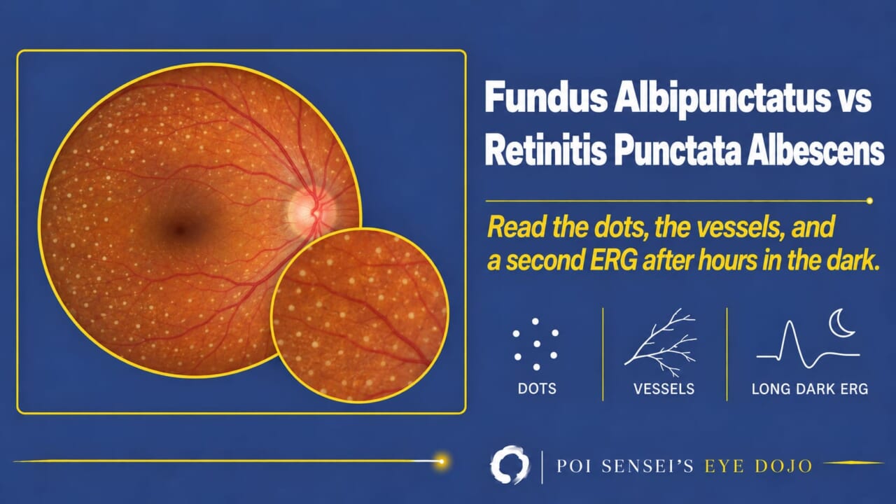
A fundus scattered with small yellow-white dots, and a patient who has not seen well in the dark since childhood. That is how both fundus albipunctatus and retinitis punctata albescens present, and the two are easy to mix up. This article shows how to separate them with the dots, the vessels and the electroretinogram recorded again after a long dark adaptation.
- Look at the dots. Are they uniform and sharp, or variable and blurred? Are the retinal vessels and the disc normal?
- Ask about the course. Has it stayed night blindness only, or is the field closing in and acuity falling?
- Record a full-field ERG after the standard dark adaptation, then again after 2 to 3 hours in the dark. Does the rod response come back?
One picture, two diseases
Fundus albipunctatus is counted among the forms of congenital stationary night blindness (CSNB) with an abnormal fundus, together with Oguchi disease[1][2]. Retinitis punctata albescens is its progressive counterpart.
The difference is in the word stationary. Fundus albipunctatus stays still in most patients, with night blindness from early life and little else. Retinitis punctata albescens progresses. The central scotoma widens, the macula atrophies, and in the end it looks like retinitis pigmentosa with white dots[1][2]. Separate them early, because the prognosis you give the family is different.
Fundus albipunctatus
Cause
Autosomal recessive in most cases, with mutations in RDH5[1][2]. RDH5 makes an enzyme the RPE needs to regenerate rhodopsin after it has been bleached by light. Without it, rhodopsin recovers very slowly. The rods do work, they just need hours instead of minutes to get ready in the dark, and once they are ready, sensitivity to dim light is normal[3].
Fundus
Countless small yellow-white dots of about the same size, with sharp edges and regular spacing, mostly in the mid-periphery and sparing the fovea[1][2]. The retinal vessels and the optic disc are normal, and there is no bone-spicule pigment[1].
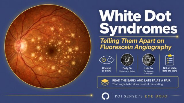
Many diseases put white dots in the fundus: retinitis punctata albescens, the white dot syndromes, drusen. Keep a picture of each in your head.
OCT
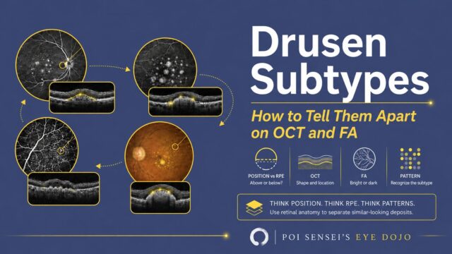
A section through a dot shows a hyperreflective deposit sitting on top of the RPE. One other deposit sits in the same place, above the RPE. Do you remember which? Subretinal drusenoid deposits, the ones that used to be called reticular pseudodrusen.
ERG
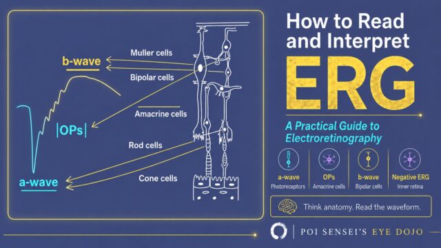
The light-adapted responses, the cone response and the 30 Hz flicker, are normal. In the dark-adapted recordings after the standard 20 to 30 minutes, the rod response is nearly flat, and the combined rod-cone response is reduced, often with a negative shape (the b-wave smaller than the a-wave) and a smaller a-wave. In other words, it looks like a negative ERG. That is the pitfall.
Keep the patient in the dark for 2 to 3 hours and record again. The rod and combined responses come back to normal or near normal[1][2].
Retinitis punctata albescens
Symptoms and course
Typically, night blindness is noticed in childhood and both fundi show white dots. Acuity and fields are relatively good in childhood, so at that age the two diseases can look alike. With age the central scotoma widens and macular atrophy appears. The course is that of retinitis pigmentosa[2].
Cause
Autosomal recessive, most often RLBP1[1]. Other genes have been reported. RLBP1 makes another protein of the same regeneration cycle, so dark adaptation is slowed here too, and far more severely[3].
Fundus
White dots again, but variable in size and less sharply demarcated, most numerous around the equator and usually sparing the macula[1]. Then look at the vessels: arteriolar attenuation is the sign fundus albipunctatus does not have[1]. Pigment may appear later in the course.
OCT
As acuity falls and the macula atrophies, OCT shows loss of the ellipsoid zone (the band that reflects photoreceptor health) and thinning of the macula.
ERG
The a- and b-waves are strongly reduced from the start, nearly flat after the standard dark adaptation. After 2 hours in the dark there is slight improvement, after 24 hours a little more, but the response does not become normal[5].
Put the two ERGs side by side
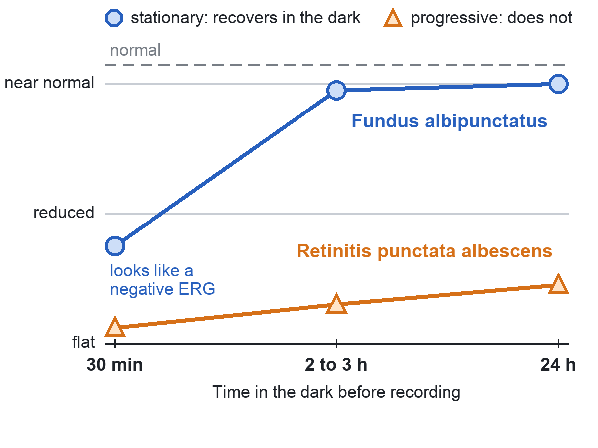
When the pictures overlap, the ERG after prolonged dark adaptation and genetic testing (RDH5 versus RLBP1) help settle it.
Summary
- Both are night blindness from childhood with a fundus full of small white dots. One stays stationary in most patients, the other progresses like retinitis pigmentosa.
- Fundus albipunctatus: uniform, sharp, regularly spaced dots, normal vessels and disc. RDH5.
- Retinitis punctata albescens: dots of varying size, arteriolar attenuation, later macular atrophy and pigment. RLBP1.
- The decisive test is the ERG after 2 to 3 hours of dark adaptation: it returns to near normal in fundus albipunctatus and stays reduced in retinitis punctata albescens.
- A fundus albipunctatus that loses acuity in later life may have a cone dystrophy on top. Test cone function with the light-adapted ERG.
References
[1] Salmon JF. Kanski’s Clinical Ophthalmology: A Systematic Approach. 10th ed. Elsevier; 2025. Chapter 15, Inherited retinal degenerations: congenital stationary night blindness.
[2] Yanoff M, Duker JS, eds. Ophthalmology. 5th ed. Elsevier; 2019. Chapter 6.14, Progressive and "stationary" inherited retinal degenerations.
[3] Levin LA, Nilsson SFE, Ver Hoeve J, Wu SM, eds. Adler’s Physiology of the Eye. 11th ed. Elsevier; 2011. Chapter 20, Box 20.2: Diseases exhibiting slowed dark adaptation.
[4] Working Group for the Clinical Practice Guideline for Retinitis Pigmentosa, Japanese Ophthalmological Society. Clinical practice guideline for retinitis pigmentosa 2026 (in Japanese). Nippon Ganka Gakkai Zasshi. 2026;130(4):376.
[5] Burstedt M, Jonsson F, Köhn L, Burstedt M, Kivitalo M, Golovleva I. Genotype-phenotype correlations in Bothnia dystrophy caused by RLBP1 gene sequence variations. Acta Ophthalmol. 2013;91(5):437–444. PMID 22551409.