Anisocoria: How to Tell Which Pupil Is Abnormal, and What to Do Next
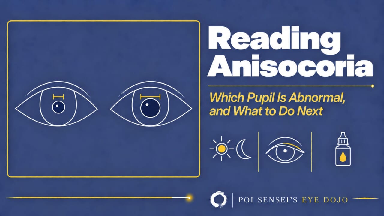
Anisocoria is one of those findings that can feel vaguely frightening. Yet few doctors work it up with real confidence. This article walks through a systematic way to find the cause of unequal pupils, one step at a time. By the end, anisocoria should no longer frighten you.
- Measure both pupils in the light and in the dark.
- Decide which pupil is abnormal: the one that fails to dilate in the dark, or the one that fails to constrict in the light.
- Look at what comes with it: ptosis, eye movements, the light reaction, the near reaction.
- Confirm with one drop, only if you still need to.
Step 1: Measure in the light and in the dark
Write down the size of each pupil in a bright room and again in a dim room, with the patient looking at a distant target. A small difference that is about the same in both conditions is physiologic anisocoria (also called simple anisocoria). It is common: in a dim room, about one in five healthy people have a difference of 0.4 mm or more at any given moment, and the difference rarely reaches 1 mm[1]. It also comes and goes: in the same study, 41% showed it at some point over five days, but only 3% showed it every time[1]. The light reaction is brisk on both sides, there is no ptosis, and eye movements are full.
Two habits help here. Measure the difference, not the absolute sizes, because the sizes change with age and lighting. And if you are unsure whether the anisocoria is old, ask for an old photograph that is close enough to see the pupils. Small ID photos are usually too grainy to help[1].
Step 2: Which pupil is abnormal?
This is the step that does the work.
- The difference is larger in the dark. The small pupil is the abnormal one. It cannot dilate, so the sympathetic pathway is the problem. Think Horner syndrome.
- The difference is larger in the light. The large pupil is the abnormal one. It cannot constrict, so the parasympathetic pathway or the iris itself is the problem. Think third nerve palsy, tonic pupil, a drug in the eye, or a damaged iris.
The logic is simple once you say it aloud: a pupil that is doing its job changes size. The one that stays put is the one that is broken.
Step 3a: The small pupil is abnormal — Horner syndrome
Horner syndrome is a lesion of the sympathetic pathway to the eye. The pupil is small, and in a dim room it dilates slowly and incompletely, so the anisocoria is most obvious in the first few seconds after the lights go down. This slow dilation is called dilation lag.
The pupil rarely comes alone. Look for:
- A mild ptosis of the upper lid, from weakness of Müller’s muscle. The lower lid is also pulled up slightly, which narrows the palpebral fissure from both sides. The eye looks sunken rather than drooping.
- Reduced sweating and flushing on the same side of the face, in some patients.
- A normal light reaction. The sphincter is intact; only dilation is slow.
The sympathetic pathway has three neurons, and the cause depends on which one is hit: the brainstem and cervical cord (first neuron, for example a lateral medullary infarct), the lung apex and neck (second neuron, for example an apical lung tumour), and the internal carotid artery up to the cavernous sinus (third neuron, for example carotid dissection or aneurysm).
Step 3b: The large pupil is abnormal
There are three common causes here, and they are told apart by what comes with the pupil.
Third nerve palsy: look at the lid and the eye movements
If the large pupil comes with ptosis and limited adduction, elevation or depression, this is a third nerve palsy, and the question becomes why. The pupillary fibres run on the surface of the nerve. A lesion that presses on the nerve from outside, such as a posterior communicating artery aneurysm, hits those fibres first. So a third nerve palsy with a dilated pupil, especially a painful one, needs urgent imaging for an aneurysm. An ischaemic palsy, as in diabetes, damages the centre of the nerve and tends to spare the pupil. This is a tendency, not a law, and the pupil can be spared early in a compressive lesion, so when in doubt, order an MRI.
Tonic pupil: look at the near reaction
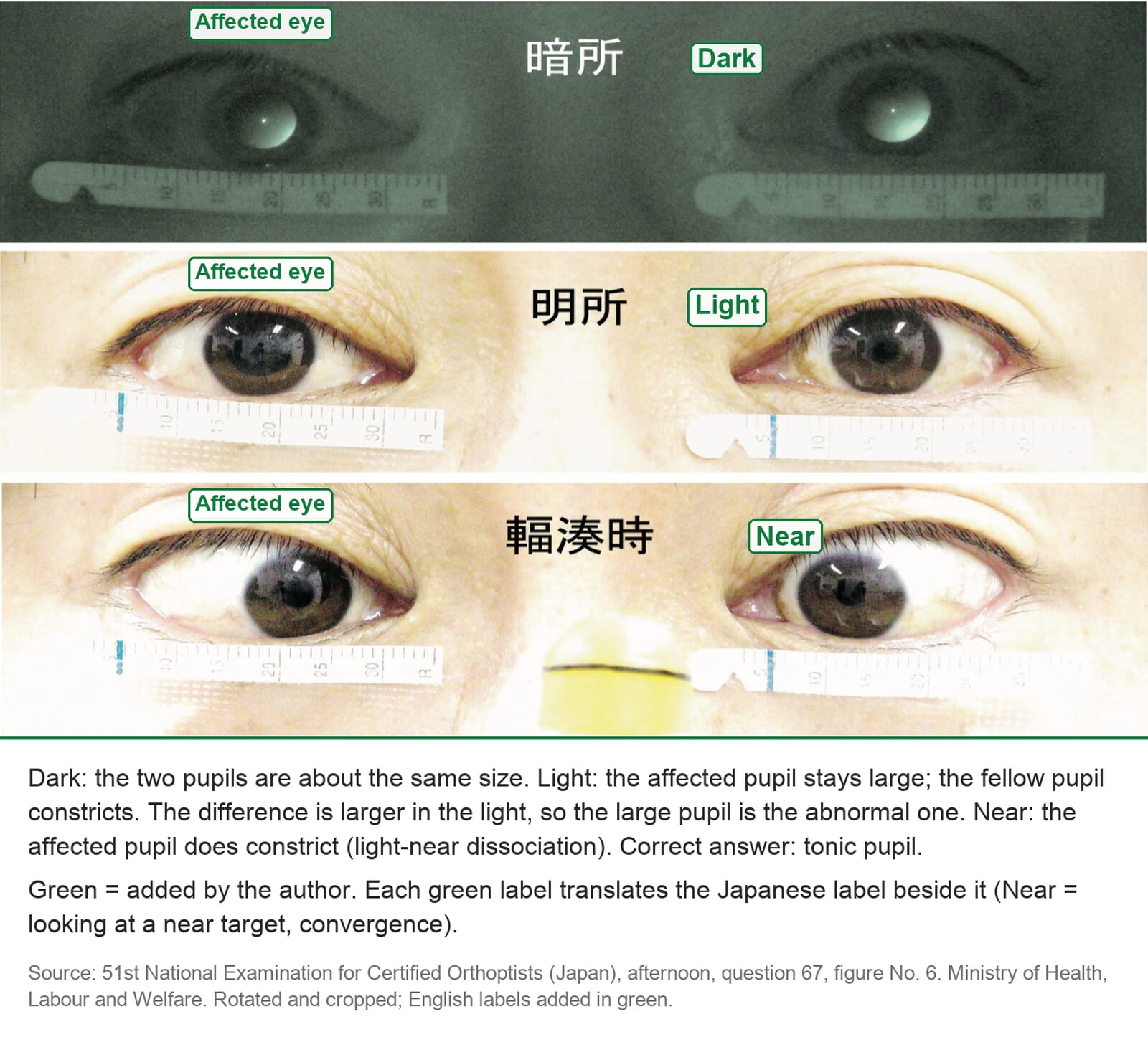
A tonic pupil (Adie pupil) is typically seen in a young adult, usually in one eye, and often found by chance or because of glare. The pupil is dilated in the light and reacts poorly to light, but it does constrict to a near target, slowly, and then stays small for a while before it redilates. This is light-near dissociation. At the slit lamp the sphincter often moves in segments rather than as a ring. In a dim room the difference shrinks, and the healthy pupil may even become the larger one, which can confuse the first examiner. If the tendon reflexes at the knee and ankle are also absent, the condition is called Adie syndrome.
Pharmacologic mydriasis and a damaged iris
A fixed, widely dilated pupil with no ptosis, full eye movements and a normal fellow eye is often a drug. Ask about scopolamine patches, nebulised anticholinergics, and plants handled in the garden. If the history is unhelpful, examine the iris at the slit lamp for sphincter tears or posterior synechiae, which point to trauma or past inflammation.
Dilute pilocarpine first (about 0.1%). If the large pupil constricts, it is a tonic pupil; the denervated sphincter is supersensitive. If it does not, move to 1% pilocarpine. If the pupil now constricts, the sphincter works and the problem is the nerve: third nerve palsy. If even 1% pilocarpine fails to constrict it, the sphincter itself is blocked or broken: a drug, or a damaged iris.
A recent cause worth asking about: sweat wipes
Glycopyrronium tosylate wipes, an anticholinergic treatment for excessive underarm sweating, are a newer cause. They were approved in the United States in 2018[3] and in Japan in 2022, and in Japan I now see this pupil more often. If the patient touches the eye or a contact lens before washing their hands, enough of the drug reaches the eye to block the sphincter. Like other drug-dilated pupils, it does not constrict even to 1% pilocarpine, and it recovers within one to two weeks once the exposure stops[2][3].
Ask for it by name. In a series of 16 patients, only 3 mentioned the wipes when asked about their medications; the rest were found only by asking specifically[2]. Patients see it as something for sweat, not as a medicine. So ask: "Do you use anything for sweating, such as wipes, creams or deodorants? Does anyone at home?"
Both pupils small with light-near dissociation
One more pattern belongs here. If both pupils are small and irregular, do not react to light, but do constrict to a near target, this is an Argyll Robertson pupil. The classic cause is neurosyphilis, which is treatable, so it is worth recognising. The lesion is thought to be near the pretectal area, on the afferent side of the light reflex.
Why an optic nerve problem does not cause anisocoria
A question I am asked often: the patient has an optic neuropathy on one side; why are the pupils equal?
Because the light reflex is consensual. Light entering either eye sends the same signal to both Edinger-Westphal nuclei, so both pupils receive the same command and stay the same size. What changes with an optic nerve lesion is the strength of the signal from that eye. That is what the swinging flashlight test measures: a relative afferent pupillary defect (RAPD), not a difference in size. The video below walks through the pathway and the test.
Summary
Measure both pupils in the light and in the dark. If the difference is larger in the dark, the small pupil is abnormal; think Horner syndrome. If the difference is larger in the light, the large pupil is abnormal; ptosis and eye movement limitation mean a third nerve palsy, light-near dissociation means a tonic pupil, and a pupil that will not constrict even to 1% pilocarpine means a drug or a damaged iris. A small difference, usually under 1 mm, that is about the same in all lighting is physiologic.
Figure credits
Photographs reproduced from the 51st National Examination for Certified Orthoptists (Japan), afternoon session, question 67, published by the Ministry of Health, Labour and Welfare. Anything in green is mine.
References
[1] Lam BL, Thompson HS, Corbett JJ. The prevalence of simple anisocoria. Am J Ophthalmol. 1987;104(1):69–73. PMID 3605282.
[2] Kaufman AR, Gulati S, Pula JH, et al. Pharmacologic mydriasis secondary to topical glycopyrronium tosylate cloths: clinical characterization from a multicenter analysis. J Neuroophthalmol. 2022;42(4):530–534. PMID 35427257.
[3] QBREXZA (glycopyrronium) cloth, 2.4%, for topical use: prescribing information. Journey Medical Corporation; revised November 2022. DailyMed, U.S. National Library of Medicine. Accessed October 6, 2026.