Optic Neuritis: Typical Features, AQP4 and MOG Antibodies, and What the ONTT Really Showed
A young woman loses vision in one eye over a few days, and it hurts when she moves the eye. That is the textbook picture of optic neuritis, and most of the time it is exactly what it looks like.
But in a Japanese nationwide study, about one in ten patients with optic neuritis carried AQP4 antibodies[1]. Their visual prognosis is poor, and they need different handling. This article goes through the typical picture, why MRI and antibody testing come first, how the treatment is decided, and what the famous Optic Neuritis Treatment Trial (ONTT) can and cannot tell you.
Features of typical optic neuritis
Typical optic neuritis is more common in relatively young women aged 15 to 45.
The symptoms are sudden visual loss in one eye and pain on eye movement.
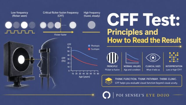
On examination, the affected eye shows a positive RAPD, a reduced critical flicker frequency (CFF), and visual field defects such as a central scotoma.
When the optic nerve is inflamed, looking up, down, left or right stretches the nerve, and that causes pain. This is why the pain comes with eye movement.
Anterior and retrobulbar optic neuritis
Optic neuritis is divided into anterior optic neuritis (papillitis), with swelling of the optic disc, and retrobulbar optic neuritis, without disc swelling.
In retrobulbar optic neuritis the fundus looks normal, so it is important to go looking for the optic nerve damage with the RAPD, the flicker test and the visual field.
MRI is essential
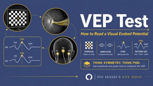
On brain MRI, STIR images show high signal in the affected optic nerve.
FLAIR images may also show demyelinating lesions around the lateral ventricles. If they are there, you need to suspect multiple sclerosis (MS).
Even when you think it is optic neuritis alone, other lesions may be hiding, and the optic neuritis may be one manifestation of MS. That is why MRI is essential.
High signal on plain MRI can also persist after treatment, so for a recurrence it is better to order contrast-enhanced MRI. (Even at the first episode, I often order contrast if renal function is good.)
Always test for antibodies
When you see optic neuritis, send anti-aquaporin-4 (AQP4) antibodies and anti-MOG antibodies.
In a Japanese nationwide survey of 531 patients with non-infectious optic neuritis, 12% were AQP4-antibody positive and 10% were MOG-antibody positive[1] — roughly one in ten each. These are Japanese data. The three groups looked like this[1]:
| AQP4 antibody positive | MOG antibody positive | Both negative | |
|---|---|---|---|
| Proportion | 12% | 10% | 77% |
| Women | 84% | 51% | 64% |
| Disc swelling | 34% | 76% | 46% |
| Pain on eye movement | 53% | 77% | 46% |
| Counting fingers or worse before treatment | 53% | 25% | 22% |
| Counting fingers or worse after treatment | 22% | 5% | 8% |
AQP4-positive optic neuritis is more common in women, and vision often fails to come back even with treatment. MOG-positive optic neuritis tends to show disc swelling and pain, but vision after treatment is good.
AQP4-positive optic neuritis has a poor visual prognosis, and it changes the treatment plan.
The MOG antibody marks a demyelinating disease that is different from both MS and AQP4-positive neuromyelitis optica: MOG antibody-associated disease (MOGAD). Besides optic neuritis, it can present as acute disseminated encephalomyelitis or myelitis, and it can be a single episode or keep relapsing[2].
MOG-positive optic neuritis has these features[2]:
- Both eyes at once is common (31–58%; under 5% in optic neuritis with MS, and 13–37% in AQP4-positive disease)
- Visual loss at onset is often severe, but it often recovers quickly with steroids
- It can relapse while steroids are being tapered or soon after they are stopped. Relapsing optic neuritis is seen in 30–50%
- MRI can show a long lesion extending along the front of the optic nerve, and enhancement of the sheath around the nerve
Treatment is steroid pulse therapy
The basic treatment is intravenous methylprednisolone (steroid pulse therapy).
The ONTT, which I introduce below, found that pulse therapy speeds up visual recovery but does not change the final visual acuity compared with no treatment. Because of this, there was a period when patients were simply observed without treatment[3].
Then the AQP4 antibody was discovered, and it became clear that positive patients should be treated early and firmly.
So in Japan, the usual flow now is:
- Send the antibody tests at admission (the results take more than a week)
- Give steroid pulse therapy while you wait for the results
- If the AQP4 antibody is positive, consider further pulse therapy or plasma exchange
When steroid pulse therapy is not enough, the next options are plasma exchange and intravenous immunoglobulin (IVIg). Where they sit differs by country:
| Plasma exchange | IVIg | |
|---|---|---|
| Japan | Covered by insurance for MS relapses that do not respond to steroids. Used for NMOSD attacks too, but not covered for NMOSD[5] | Approved in 2019 for the acute phase of optic neuritis when steroids are not effective enough, in principle for AQP4-positive patients[4][5] |
| England (NHS) | The step before IVIg[6] | Funded for attacks that do not respond to IV methylprednisolone and plasma exchange, or when plasma exchange is not available, delayed or contraindicated[6] |
| German-speaking countries (NEMOS recommendations) | Start early if the response to steroids is insufficient within the first days. Can be the first choice for severe attacks[7] | Not a standard attack treatment. An option mainly for children, for patients who cannot have other treatments, or as an add-on[7] |
Optic neuritis can recur or progress to MS, so you still need to be careful in follow-up after treatment.
A famous trial: the ONTT
The trial you cannot skip when you learn about optic neuritis is the Optic Neuritis Treatment Trial (ONTT)[3].
The ONTT randomly assigned 457 patients with acute optic neuritis to three groups:
- Oral prednisone 1 mg/kg/day for 14 days
- Intravenous methylprednisolone 250 mg every 6 hours for 3 days (1 g/day), followed by oral prednisone 1 mg/kg/day for 11 days
- Oral placebo for 14 days
Patients were aged 18 to 46, with acute optic neuritis in one eye within 8 days of symptom onset, and with an RAPD and a visual field defect. The mean age was 32, and 77% were women.
The results:
- With pulse therapy, recovery was significantly faster, especially in the visual field
- At 6 months, the pulse group was slightly better in visual field, contrast sensitivity and colour vision, but visual acuity did not differ between the three groups
- Oral prednisone alone gave no better recovery than placebo, and optic neuritis actually recurred more often (relative risk 1.79)
- During follow-up (6 to 24 months), the proportion diagnosed with MS in either steroid group did not differ significantly from placebo
The ONTT also looked at many other things, such as what symptoms and visual field patterns the patients had, and what the risk factors for developing MS were. If you are interested, read the original paper.
Limits of the ONTT
However, the ONTT included only patients with one eye affected. Patients with both eyes affected were excluded. The upper age limit was 46, so older patients were not included either.
This means the ONTT may have included almost none of the optic neuritis with a poor prognosis, such as AQP4-positive disease.
So "the ONTT showed no difference in visual acuity, so no treatment is fine" does not follow. As I wrote above, I think the safe approach is to give steroid pulse therapy while you wait for the antibody results.
The ONTT is an old paper, but it is very useful for learning about optic neuritis.
Every field of ophthalmology has famous studies that come up again and again, and textbook summaries often drift away from what the paper actually said. I recommend getting into the habit of reading the original.
Summary
- Sudden visual loss in one eye with pain on eye movement in a young woman, with a positive RAPD and a reduced CFF, is typical optic neuritis.
- Even when it looks typical, do not forget to test for AQP4 and MOG antibodies.
- Treatment is steroid pulse therapy. If it is not effective enough, other treatments may be needed depending on the antibody results.
References
[1] Ishikawa H, Kezuka T, Shikishima K, et al. Epidemiologic and Clinical Characteristics of Optic Neuritis in Japan. Ophthalmology. 2019;126(10):1385–1398. PMID 31196727.
[2] Banwell B, Bennett JL, Marignier R, et al. Diagnosis of myelin oligodendrocyte glycoprotein antibody-associated disease: International MOGAD Panel proposed criteria. Lancet Neurol. 2023;22(3):268–282. PMID 36706773.
[3] Beck RW, Cleary PA, Anderson MM, et al. A randomized, controlled trial of corticosteroids in the treatment of acute optic neuritis. The Optic Neuritis Study Group. N Engl J Med. 1992;326(9):581–588. PMID 1734247.
[4] Teijin Pharma. Press release on the additional indication for Kenketsu Venilon-I (in Japanese). 20 December 2019. https://www.teijin-pharma.co.jp/pressrelease/2019/20191220.html
[5] Japanese Society of Neurology. Clinical practice guidelines for multiple sclerosis and neuromyelitis optica spectrum disorder 2023 (in Japanese). Igaku-Shoin; 2023. pp.102–103 (insurance coverage) and the sections on plasmapheresis and IVIg. https://www.neurology-jp.org/guidelinem/pdf/koukasyo_nmosd_2023.pdf
[6] NHS England. Clinical Commissioning Policy for the use of therapeutic immunoglobulin (Ig) England (2024), version 2.0. April 2024. Section on non-MS CNS inflammatory disease (NMOSD, MOGAD, optic neuritis). https://igd.mdsas.com/wp-content/uploads/ccp-for-the-use-of-therapeutic-immunoglobulin-england-2024-v4.pdf
[7] Kümpfel T, Giglhuber K, Aktas O, et al. Update on the diagnosis and treatment of neuromyelitis optica spectrum disorders (NMOSD) – revised recommendations of the Neuromyelitis Optica Study Group (NEMOS). Part II: Attack therapy and long-term management. J Neurol. 271(1):141–176 (published online 2023). PMID 37676297.