Enhanced S-Cone Syndrome: The ERG Pattern That Makes the Diagnosis
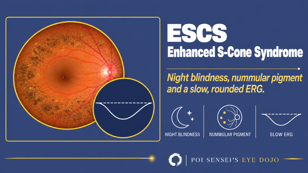
A child has night blindness, and the fundus shows round pigment clumps along the vascular arcades. It looks like retinitis pigmentosa. But the ERG has a shape you see in no other disease. That is enhanced S-cone syndrome (ESCS). This article explains what ESCS is, and how to read the ERG that makes the diagnosis.
- Night blindness has been there since early childhood.
- Round pigment clumps or yellow-white dots sit along the vascular arcades.
- The macula has cystic spaces, but they do not leak on fluorescein angiography.
- The rod response is absent, and the mixed response (normally rods and cones together) and the cone response look the same: slow and rounded.
What is enhanced S-cone syndrome?
The retina has three kinds of cones. S-cones respond to short-wavelength (blue) light, M-cones to medium and L-cones to long wavelengths. In most retinal dystrophies, all of them get weaker.
ESCS is one of the few in which one type of photoreceptor, the S-cone, works more than normal. The rods do not work, and the M- and L-cones are weak[1].
It is autosomal recessive, caused by mutations in NR2E3. Goldmann-Favre syndrome is a severe form of the same disease[1].
Symptoms
Night blindness is the first symptom in most patients, usually in early childhood[3]. Vision varies widely, from worse than 20/200 to 20/20. Hyperopia is common[3].
Color vision is reduced, but blue-yellow discrimination is relatively spared, because the S-cones are working[1].
Fundus and OCT
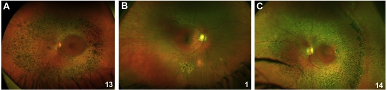
The typical sign is nummular (coin-shaped) pigment clumps along the vascular arcades and in the mid-periphery. Yellow-white dots are also common[3]. The fundus can also look almost normal.
A fundus like this is easily called retinitis pigmentosa. An ERG is what tells them apart.
On OCT, the macula often shows cystic spaces or schisis (splitting of the retinal layers). Unlike cystoid macular edema, they do not leak on fluorescein angiography[1]. The ellipsoid zone may be hard to see.
ERG findings
The ERG pattern of ESCS is so typical that it is enough to make the diagnosis[4]. Start with the mixed response (DA 3.0), and compare it with a normal one.
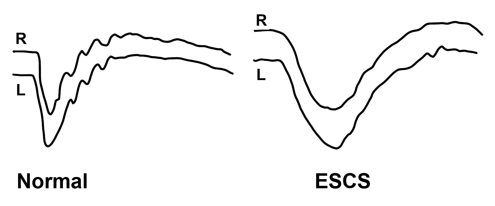
The mixed response is slow and rounded
In the normal response, the a-wave falls sharply, and the b-wave rises fast with small wavelets on it. In ESCS, the a-wave falls slowly, the trough is wide and round, and the b-wave climbs back slowly. The whole wave is slow and simple[4].
The a-wave is delayed most of all. In my practice, I call the a-wave of the mixed response delayed when its peak time is over 20 ms.
The other steps
DA means dark-adapted and LA means light-adapted. The number is the flash strength.
| ERG step | ESCS |
|---|---|
| Rod (DA 0.01) | Absent |
| Mixed response (DA 3.0) | Slow and rounded |
| Cone (LA 3.0) | Same shape as the mixed response |
| 30 Hz flicker | Smaller than the cone a-wave |
| S-cone ERG | Enlarged |
The rods do not work, so the rod response cannot be recorded[1].
The cone response has the same slow, rounded shape as the mixed response, because in the dark and in the light the response comes mainly from S-cones[2].
Normally, the 30 Hz flicker is larger than the a-wave of the cone response and smaller than its b-wave. In ESCS it is smaller than the a-wave[3][4]. S-cones are slow and cannot follow a fast flicker well.
A blue flash on an orange background records the S-cones on their own (the S-cone ERG). In ESCS this response is much larger than normal[2]. This test is not available everywhere. The standard ERG is usually enough.
Treatment and prognosis
There is no cure. Compared with retinitis pigmentosa, it progresses slowly. In a series of 56 patients, vision stayed stable in most eyes over about 6 years[3].
Summary
- ESCS is an autosomal recessive disease (NR2E3) in which S-cones are overactive, rods do not work, and M- and L-cones are weak.
- Night blindness starts in early childhood. Nummular pigment along the arcades and macular cysts without leakage are typical.
- ERG: no rod response, a mixed response and cone response with the same slow shape, and a 30 Hz flicker smaller than the cone a-wave.
- It progresses slowly. Separate it from retinitis pigmentosa with the ERG.
Figure credits
Fundus photographs: Figure 2 (panels A to C) from de Carvalho ER, Robson AG, Arno G, Boon CJF, Webster AA, Michaelides M. Ophthalmol Retina 2021;5(2):195–214 (https://doi.org/10.1016/j.oret.2020.07.008), licensed under CC BY 4.0. Figure 2 cropped to panels A to C. The ERG drawing is my own.
References
[1] Salmon JF. Kanski’s Clinical Ophthalmology: A Systematic Approach. 10th ed. Elsevier; 2025. Chapter 15, Inherited retinal degenerations: Enhanced S-cone syndrome and Goldmann-Favre syndrome.
[2] Yanoff M, Duker JS, eds. Ophthalmology. 5th ed. Elsevier; 2019. Chapter 6.9, Retinal electrophysiology.
[3] de Carvalho ER, Robson AG, Arno G, Boon CJF, Webster AA, Michaelides M. Enhanced S-cone syndrome: spectrum of clinical, imaging, electrophysiologic, and genetic findings in a retrospective case series of 56 patients. Ophthalmol Retina. 2021;5(2):195–214. PMID 32679203.
[4] Robson AG, Nilsson J, Li S, et al. ISCEV guide to visual electrodiagnostic procedures. Doc Ophthalmol. 2018;136(1):1–26. PMID 29397523.
Which retinal layer makes which wave, the three abnormal patterns, and the dark-adaptation trap to check before you report “negative” — on one page, by email. One email when I publish something new, about once a month. Unsubscribe anytime.