X-Linked Retinoschisis: Spoke-Wheel Macula, OCT and Negative ERG
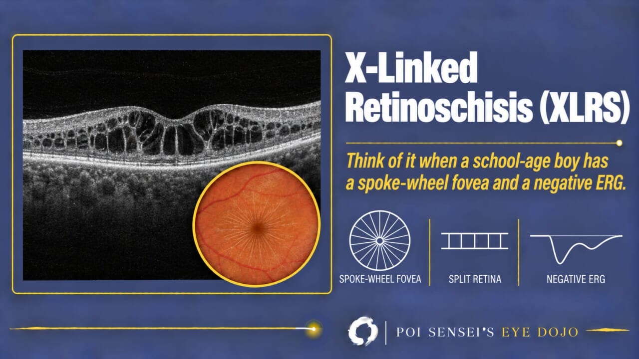
A boy around school age has poor vision in both eyes, often with hyperopia, and his fovea looks like the spokes of a bicycle wheel. That is the typical picture of X-linked retinoschisis (XLRS). This article shows how to read the fundus, the optical coherence tomography (OCT) and the electroretinogram (ERG), and how schisis differs from a retinal detachment.
- A boy has reduced vision in both eyes from around school age.
- The fovea shows fine radial folds, like the spokes of a wheel.
- OCT shows the retina split into layers in both eyes.
- The ERG is electronegative (the b-wave is smaller than the a-wave).
What is X-linked retinoschisis?
XLRS is a congenital disease in which the retina splits within its own layers. The split is at the macula and sometimes also in the periphery. It is also called congenital or juvenile retinoschisis.
Inheritance is X-linked recessive, so XLRS almost always affects boys. Carrier mothers usually have no symptoms and a normal fundus[1].
The gene is RS1, named after RetinoSchisis. It makes retinoschisin, a protein that helps retinal cells hold together. Few inherited retinal diseases are X-linked, so XLRS is worth remembering. Choroideremia is another one. XLRS affects about 1 in 5,000 to 20,000 people[2].
Symptoms
Most boys come in around school age, between 5 and 10 years. They have poor vision, trouble reading, amblyopia or strabismus. Hyperopia is common, and the amblyopia is often hyperopic[2].
Vision is usually only moderately reduced, often around 20/100 to 20/30. Later in adult life it tends to fall as the macula becomes atrophic[3].
Less often, XLRS is found in infancy because of strabismus or nystagmus. These babies often have severe peripheral schisis and vitreous hemorrhage.
Examination findings
Fundus
The key sign is foveal schisis: fine folds radiating from the fovea, like the spokes of a bicycle wheel. It is almost always in both eyes. In adults the spokes fade, and the macula may look simply atrophic.
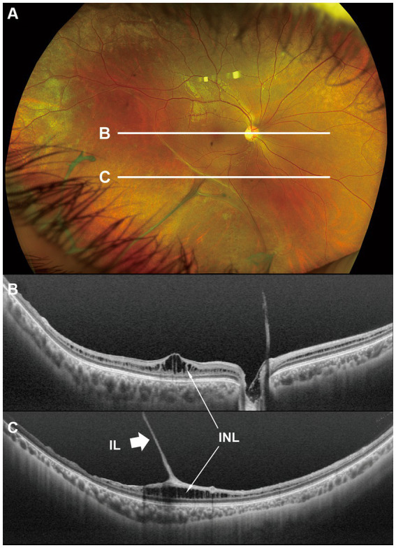
About half of patients also have peripheral schisis, most often in the inferotemporal retina. The inner wall of the split is very thin. Large oval holes can form in it, sometimes leaving only retinal vessels floating in the vitreous ("vitreous veils").
Some eyes show a silver-gray metallic sheen in the peripheral retina, like the sheen of Oguchi disease[2].
OCT
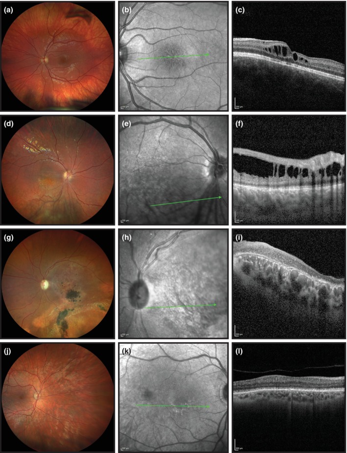
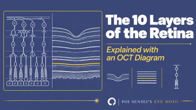
OCT shows which retinal layers are split. At the macula, the split shows up as cyst-like spaces, mainly in the inner nuclear layer and outer plexiform layer[1]. In the periphery, the thin inner layer lifts into the vitreous.
Compare this with a retinal detachment. In a detachment, the retina separates between the photoreceptors and the retinal pigment epithelium (RPE). In schisis, the photoreceptors stay on the RPE, and the split is inside the retina.
ERG
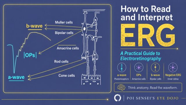
The typical ERG is electronegative. The a-wave comes from the photoreceptors, and the b-wave mainly from the bipolar cells. In the dark-adapted bright flash, the a-wave is normal but the b-wave is smaller than the a-wave. The photoreceptors are working, but the signal passing to the inner retina is impaired. The electronegative ERG article explains the waveform.
The full-field ERG reflects the whole retina, so an eye with only macular schisis can have a normal ERG[1]. A normal ERG does not rule out XLRS. Genetic testing for RS1 confirms the diagnosis.
Treatment
There is no established treatment that cures XLRS.
- In children, correct the refractive error, treat amblyopia, and check often for vitreous hemorrhage and retinal detachment.
- Carbonic anhydrase inhibitors (dorzolamide drops or oral acetazolamide) can shrink the foveal cysts and improve vision in some patients. Results vary between studies[3].
- Vitrectomy may be needed for vitreous hemorrhage or retinal detachment[1].
- Gene therapy is still under investigation.
Summary
- XLRS is an X-linked disease caused by RS1, seen almost only in boys.
- Look for a spoke-wheel fovea in both eyes and inferotemporal peripheral schisis.
- OCT shows the split inside the retina, mainly in the inner nuclear and outer plexiform layers. In a detachment, the split is under the photoreceptors.
- The ERG is typically electronegative, but it can be normal in eyes with only macular schisis.
- In children, correct hyperopia, treat amblyopia, and watch for hemorrhage and detachment.
Figure credits
Fundus photographs and OCT at different ages: Figure 1 from Järvinen MA, Baraas RC, Majander A, et al. Acta Ophthalmol 2025;103:196-204, licensed under CC BY 4.0. Unchanged.
Peripheral schisis: Figure 1 from Nakajima A, Kuniyoshi K, Iwahashi C, et al. Front Med (Lausanne) 2023;10:1280564 (https://doi.org/10.3389/fmed.2023.1280564), licensed under the Creative Commons Attribution License (CC BY). Unchanged.
References
[1] Salmon JF. Kanski’s Clinical Ophthalmology: A Systematic Approach. 10th ed. Elsevier; 2025. Chapter 15, Inherited retinal degenerations: Juvenile X-linked retinoschisis.
[2] Nakajima A, Kuniyoshi K, Iwahashi C, et al. Optical coherence tomography findings of the peripheral retina in patients with congenital X-linked retinoschisis. Front Med (Lausanne). 2023;10:1280564. PMID 38034549.
[3] Järvinen MA, Baraas RC, Majander A, et al. A retrospective longitudinal study of 52 Finnish patients with X-linked retinoschisis. Acta Ophthalmol. 2025;103(2):196-204. PMID 39435478.
Which retinal layer makes which wave, the three abnormal patterns, and the dark-adaptation trap to check before you report “negative” — on one page, by email. One email when I publish something new, about once a month. Unsubscribe anytime.