Gyrate Atrophy of the Choroid and Retina: Fundus, Ornithine and Treatment
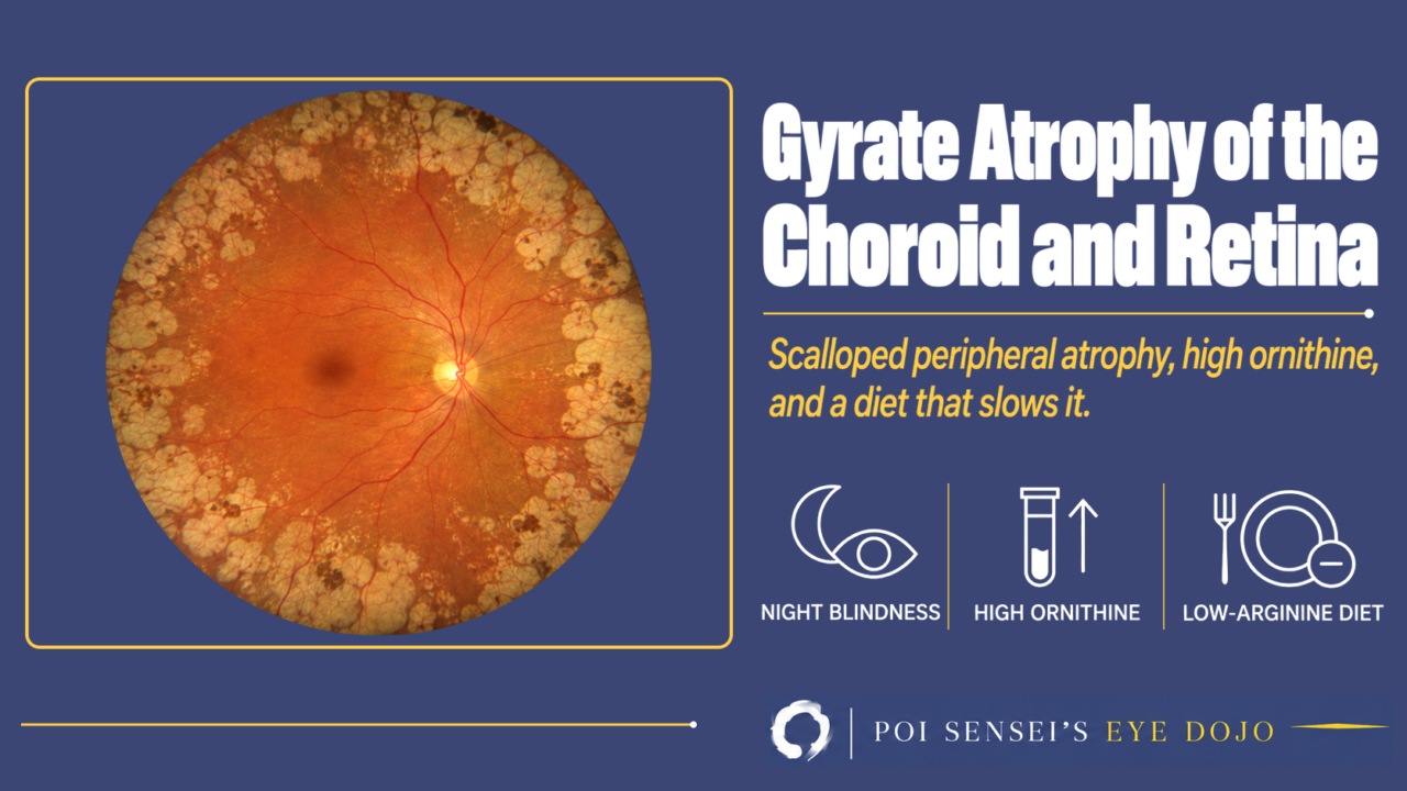
Sharply demarcated, scalloped patches of atrophy in the mid-periphery, night blindness and high myopia. Think of gyrate atrophy. This article shows how to recognize it, what the electroretinogram (ERG) and OCT show, how a blood test confirms it, and what treatment can do.
- Night blindness starts in childhood or the teens, with high myopia.
- The mid-peripheral fundus shows round, sharply demarcated patches of chorioretinal atrophy that join together.
- The macula is spared until late.
- Posterior subcapsular cataract appears at a young age.
What is gyrate atrophy?
Gyrate atrophy of the choroid and retina is an autosomal recessive disease. Mutations in the OAT gene make the enzyme ornithine aminotransferase (OAT) work poorly, so ornithine builds up in the blood.
"Gyrate" means shaped like the gyri, the folds on the surface of the brain. The joined patches of atrophy in the fundus look like these folds.
Symptoms
The first symptom is night blindness, usually in childhood or the teens. Some patients are not diagnosed until adulthood.
As the choroid and retina waste away, the visual field narrows from the periphery. Central vision is kept until late. The field keeps narrowing, and many patients become legally blind around the age of 50[1].
Most patients are myopic, often highly myopic. Posterior subcapsular cataract is common and starts young. It can lower vision, and so can cystoid macular edema.
Findings
Fundus
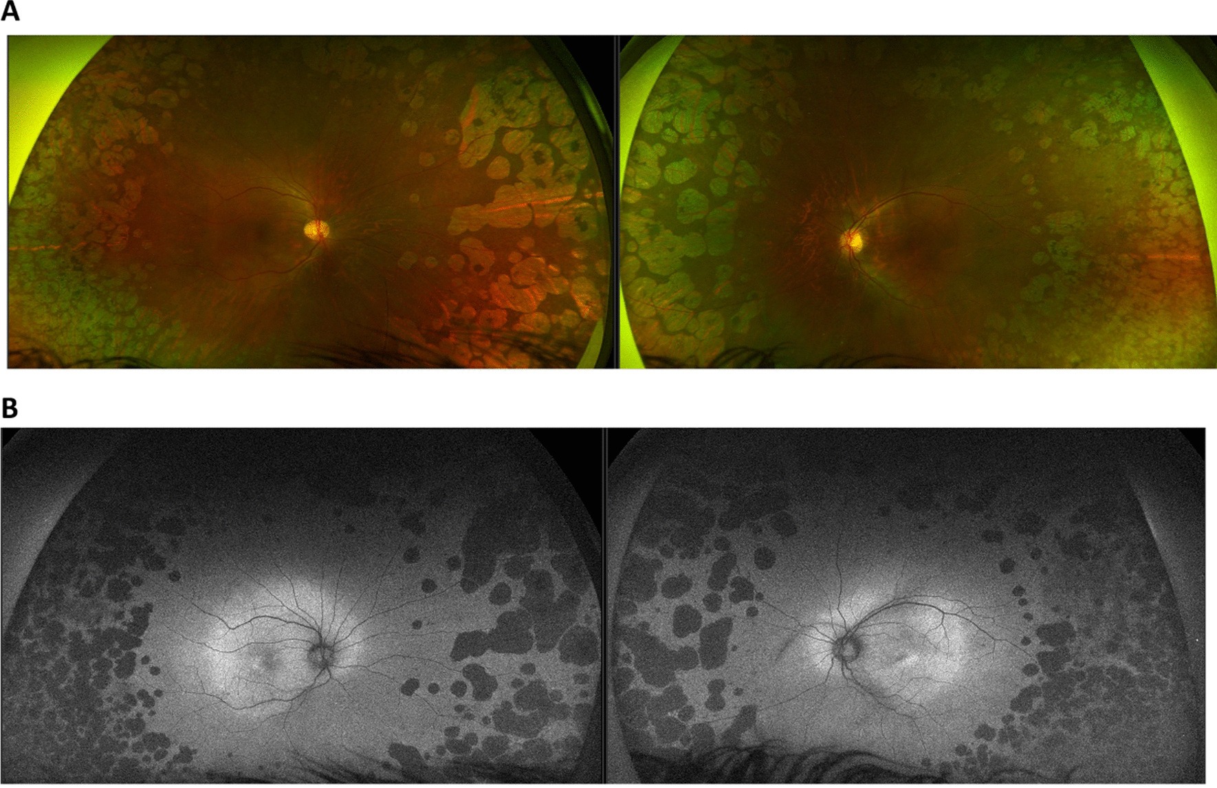
The fundus shows round or scalloped (wavy-edged) patches of chorioretinal atrophy with sharp borders. They start in the mid-periphery, grow and join together, then spread toward the posterior pole and the far periphery. Through the atrophy you can see the large choroidal vessels. The fovea is spared until late.
OCT
OCT shows thinning of the outer retina and the retinal pigment epithelium (RPE), with loss of the ellipsoid zone (EZ), the line made by the photoreceptors. Cystoid macular edema is also common, seen in about one third of patients in one series[2].
ERG
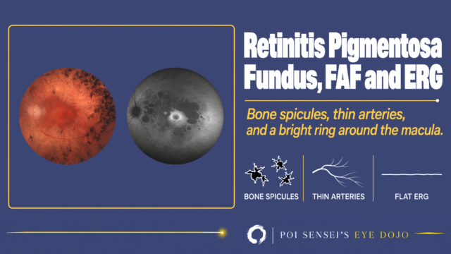
The ERG is reduced from the early stage. Rod function is lost first, then cone function. In advanced disease the ERG shows almost no response.
EOG
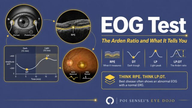
The electro-oculogram (EOG) is a test of RPE function. The RPE becomes thin over a wide area, so its result, the light peak to dark trough (LP:DT) ratio, falls as the disease progresses[3].
Diagnosis
Measure ornithine in the blood. Plasma ornithine is raised, often to 10 to 20 times normal, and urine ornithine is also high[3]. Genetic testing finds mutations in both copies of OAT.
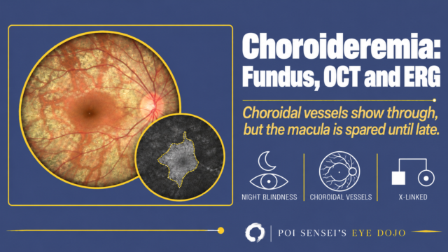
In late stages the fundus can look like choroideremia. Choroideremia is X-linked, and its ornithine level is normal.
Why ornithine builds up
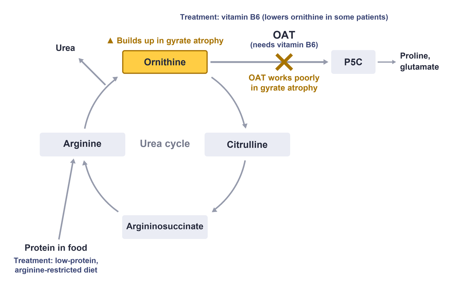
Ornithine is part of the urea cycle. Normally OAT breaks it down, and OAT needs vitamin B6 to work. When OAT works poorly, ornithine is not broken down and builds up. Ornithine is made mainly from arginine, which comes from protein in the diet.
Treatment
There is no cure. High ornithine is linked to damage of the retina, so treatment aims to lower it.
A low-protein, arginine-restricted diet lowers ornithine and can slow the disease. The diet is very strict, though, and many patients find it hard to keep[1].
Vitamin B6 lowers ornithine in only a small minority of patients, who tend to have milder disease. You cannot tell in advance who will respond, so every newly diagnosed patient can be given a trial of B6[2].
Cataract surgery may improve vision for a while.
Summary
- Gyrate atrophy is an autosomal recessive disease caused by OAT deficiency, with high ornithine in the blood.
- It starts with night blindness and high myopia, and the visual field narrows from the periphery.
- Vision can also drop from cystoid macular edema or cataract.
- The fundus shows sharply demarcated, scalloped patches of atrophy in the mid-periphery that join together. The fovea is spared until late.
- The ERG is reduced early and shows almost no response later. The EOG ratio falls as the RPE thins.
- A plasma ornithine level confirms it. A low-protein, arginine-restricted diet can slow the disease, and some patients respond to vitamin B6.
Figure credits
Fundus photographs and autofluorescence images: Figure 1A and 1B from Palmer E, Stepien KM, Campbell C, et al. Clinical, biochemical and molecular analysis in a cohort of individuals with gyrate atrophy. Orphanet J Rare Dis 2023;18:265, licensed under CC BY 4.0. Cropped (panel C removed).
References
[1] Salmon JF. Kanski’s Clinical Ophthalmology: A Systematic Approach. 10th ed. Elsevier; 2025. Chapter 15, Inherited retinal degenerations: Gyrate atrophy.
[2] Palmer E, Stepien KM, Campbell C, Barton S, Iosifidis C, Ghosh A, Broomfield A, Woodall A, Wilcox G, Sergouniotis PI, Black GC. Clinical, biochemical and molecular analysis in a cohort of individuals with gyrate atrophy. Orphanet J Rare Dis. 2023;18(1):265. PMID 37667371.
[3] Yanoff M, Duker JS, eds. Ophthalmology. 5th ed. Elsevier; 2019. Chapter 6.16, Choroidal dystrophies: Gyrate atrophy.
Which retinal layer makes which wave, the three abnormal patterns, and the dark-adaptation trap to check before you report “negative” — on one page, by email. One email when I publish something new, about once a month. Unsubscribe anytime.