Oguchi Disease: The Golden Fundus, the Mizuo-Nakamura Phenomenon and the ERG
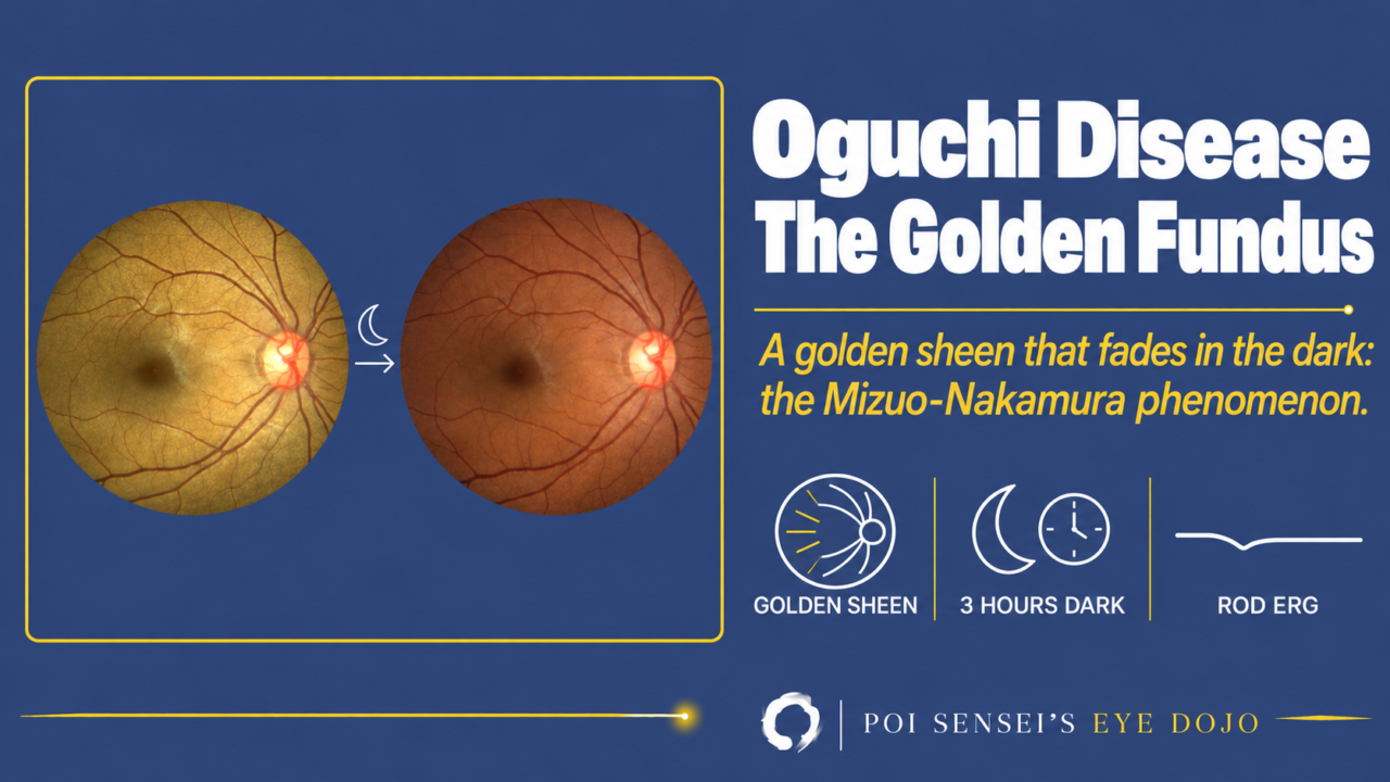
A young patient has a golden, metallic-looking fundus and has had trouble seeing in the dark since childhood. Think of Oguchi disease. This article explains how to confirm it with the Mizuo-Nakamura phenomenon and the electroretinogram (ERG), and how to tell it from fundus albipunctatus.
- The fundus has a golden-yellow metallic sheen.
- The sheen fades after a few hours in the dark.
- Night blindness is often mild and usually does not get worse. Visual acuity is normal.
- After routine dark adaptation, the rod response is absent, but the cone responses are normal.
What is Oguchi disease?
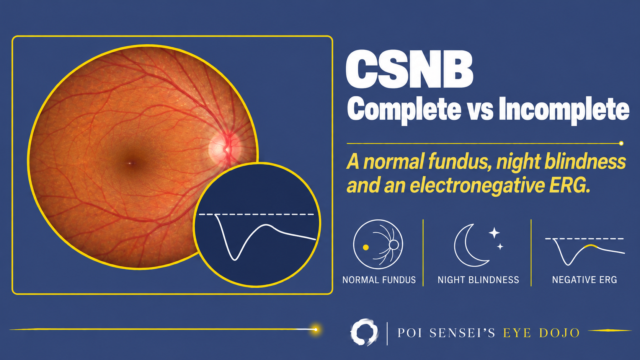
Oguchi disease is one form of congenital stationary night blindness (CSNB). Night blindness starts in childhood and usually does not progress. Unlike CSNB with a normal fundus, Oguchi disease has a characteristic fundus appearance.
It was first described in 1907 by the Japanese ophthalmologist Chuta Oguchi.
Symptoms
Typical patients notice night blindness from a young age. But many have only mild symptoms, so the disease is often found by chance when someone looks at the fundus. Visual acuity, visual fields and color vision are usually normal[1].
Inheritance
Oguchi disease is autosomal recessive. Two genes are known: SAG, which codes for arrestin, and GRK1, which codes for rhodopsin kinase. Both proteins switch off rhodopsin after it has absorbed light[2].
When either protein fails, the rods take hours to recover their sensitivity after light. Cone function is normal.
Findings
Fundus
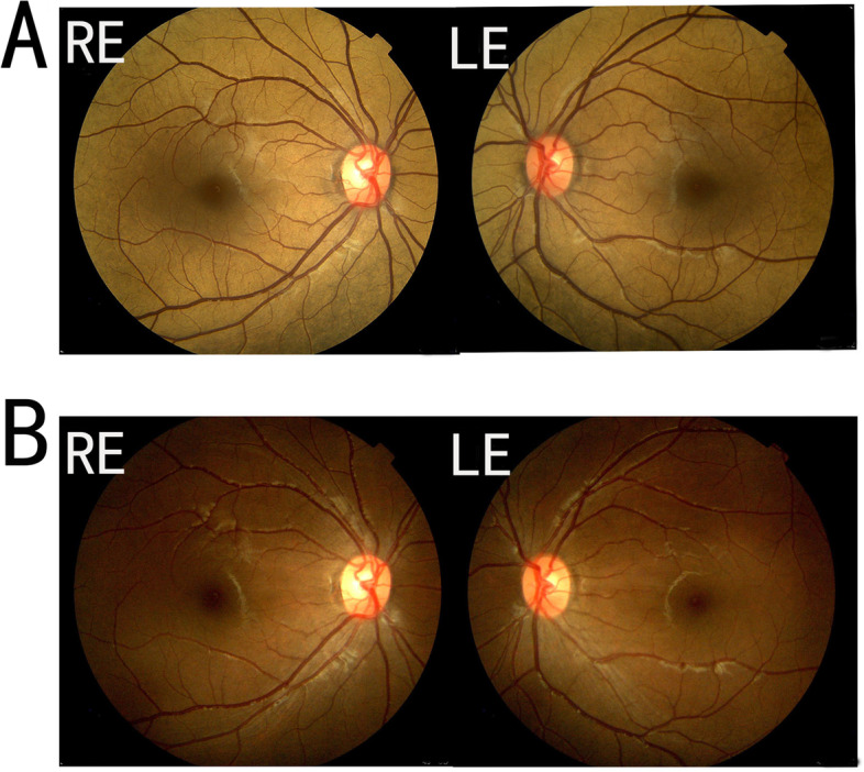
The fundus shows a golden-yellow metallic sheen (a tapetal-like reflex). How much of the fundus shows it varies a lot between patients. It may cover the whole fundus or only part of it, and some eyes also show areas of degeneration.
After about 3 hours of dark adaptation, the sheen disappears and the fundus looks normal. This is the Mizuo-Nakamura phenomenon. When the eye is exposed to light again, the sheen comes back[2].
ERG
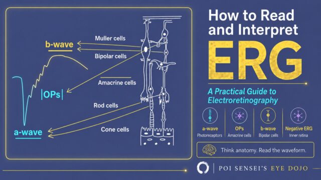
After the routine dark adaptation (20 to 30 minutes), the rod response is absent. In the mixed response (dark-adapted 3.0 ERG), the a-wave is reduced and the b-wave is smaller than the a-wave (an electronegative waveform). The cone response and the 30 Hz flicker (also a cone test) are normal[2].
This looks the same as a normal eye recorded with too little dark adaptation. In Oguchi disease, the rods need far more than the routine 20 to 30 minutes.
With very long dark adaptation, the rod response improves. But rod function recovers more slowly than the fundus color, and the ERG often stays smaller than normal[2].
Treatment
No treatment is needed. Vision and visual fields usually stay good.
But Oguchi disease is not always stationary. Some patients develop retinal degeneration in adulthood, and a few go on to retinitis pigmentosa. Do not promise patients that it will never change[4].
Oguchi disease vs fundus albipunctatus
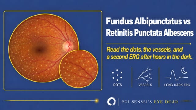
Both cause stationary night blindness with an abnormal fundus. In both, the rods recover after long dark adaptation. The fundus tells them apart.
| Oguchi disease | Fundus albipunctatus | |
|---|---|---|
| Fundus | Golden metallic sheen | Many small white dots |
| Where | Whole fundus or patches | Spares the fovea |
| In the dark | Sheen fades | Dots stay |
Read more in fundus albipunctatus vs retinitis punctata albescens.
Summary
- Oguchi disease is an autosomal recessive form of congenital stationary night blindness, caused by SAG or GRK1.
- The fundus has a golden metallic sheen that disappears after about 3 hours in the dark (Mizuo-Nakamura phenomenon).
- After routine dark adaptation, the rod response is absent and the mixed response is electronegative. Cone responses are normal.
- With very long dark adaptation, the rods improve, but the ERG often stays smaller than normal.
- No treatment is needed. It is usually stationary, but some patients develop retinal degeneration in adulthood.
Figure credits
Mizuo-Nakamura phenomenon: Figure 1 from Deng Z, Fan F, Tang D, Wu Y, Shu Y, Wu K. BMC Ophthalmol 2022;22:99 (https://doi.org/10.1186/s12886-022-02307-z), licensed under CC BY 4.0. Unchanged.
References
[1] Deng Z, Fan F, Tang D, Wu Y, Shu Y, Wu K. A compound heterozygous mutation in the S-Antigen Visual Arrestin SAG gene in a Chinese patient with Oguchi type one: a case report. BMC Ophthalmol. 2022;22(1):99. PMID 35246075.
[2] Yanoff M, Duker JS, eds. Ophthalmology. 5th ed. Elsevier; 2019. Chapter 6.14, Progressive and "stationary" inherited retinal degenerations.
[3] Mirshahi A, Hassanpoor N, Khojasteh H, Baradaran MR, Faghihi H, Lashay A. Oguchi disease associated with keratoconus. J Ophthalmic Vis Res. 2021;16(1):137-139. PMID 33520139.
[4] Nishiguchi KM, Ikeda Y, Fujita K, et al. Phenotypic features of Oguchi disease and retinitis pigmentosa in patients with S-antigen mutations: a long-term follow-up study. Ophthalmology. 2019;126(11):1557-1566. PMID 31257036.
Which retinal layer makes which wave, the three abnormal patterns, and the dark-adaptation trap to check before you report “negative” — on one page, by email. One email when I publish something new, about once a month. Unsubscribe anytime.