Stargardt Disease: Flecks, Autofluorescence, the Dark Choroid and ERG Groups
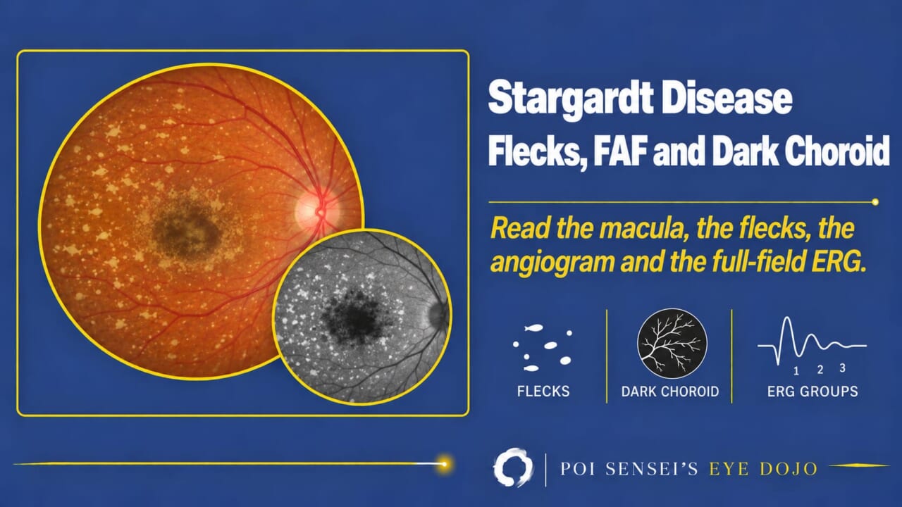
A young patient with vision falling in both eyes, and a macula that looks atrophic or, early on, almost normal. Stargardt disease is the first inherited cause to think of. This article shows how to read the fundus, autofluorescence, angiogram and ERG in Stargardt disease, and why the choroid goes dark.
- Look for macular atrophy and yellow flecks around it.
- Take a fundus autofluorescence image. Is the atrophy dark and are the flecks bright?
- If an angiogram has been done, is the background choroid dark?
- Record a full-field ERG to see how far beyond the macula the disease has spread.
What Stargardt disease is
Stargardt disease is the most common inherited macular dystrophy[1][2]. Most cases are autosomal recessive, with mutations in ABCA4[1][2].
You will also see the name fundus flavimaculatus, for the form with flecks spread beyond the macula. It is now regarded as part of the same disease[1].
Symptoms
Onset is usually in the first two decades[2]. Patients notice slowly progressive loss of vision in both eyes, a central field defect and photophobia.
Because it is common, put Stargardt disease on the list whenever a young patient has bilateral macular atrophy and falling acuity.
Findings
Fundus
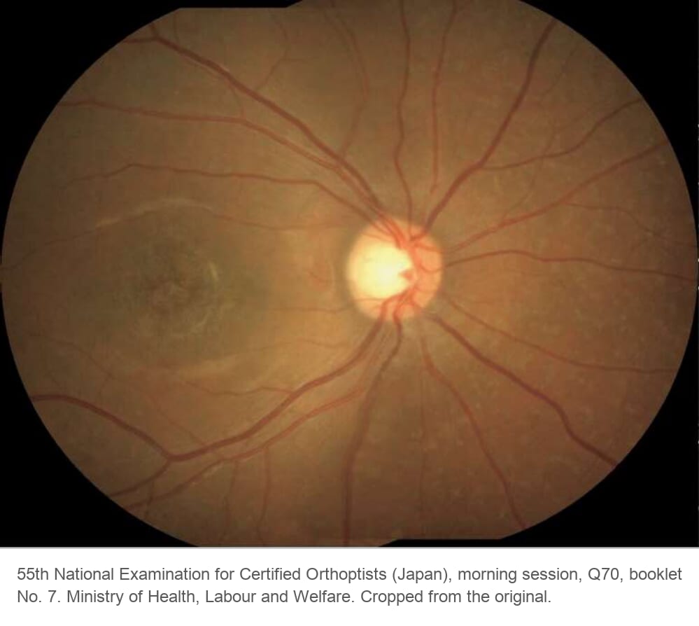
The two signs to look for are macular atrophy and yellow flecks around it[1][2]. The flecks are yellow-white, round, oval or pisciform (fish-shaped), and lie at the level of the retinal pigment epithelium (RPE). They may stay in the posterior pole or extend to the mid-periphery[1].
Severity and appearance vary widely, even within one family[2]. Do not expect every patient to look like the textbook picture.
Fundus autofluorescence
Lipofuscin, a waste product of the retina’s metabolism, builds up in the RPE and autofluoresces. That makes fundus autofluorescence the key test. The atrophic macula is dark, the flecks are usually bright, and the background is often a little brighter than normal[1][3]. The retina around the optic disc tends to be spared (peripapillary sparing), which supports the diagnosis[1][3].
Fluorescein angiography: the dark choroid
Normally the dye fills the choroid behind the retina and gives the background of the angiogram a faint glow (the window effect). In Stargardt disease this glow is missing, and the retinal vessels stand out against a dark background. This is the dark choroid[1][2].
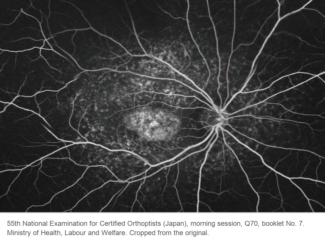
Today autofluorescence and OCT have largely replaced the angiogram for this diagnosis[1]. Not every patient shows a dark choroid, so its absence does not rule out the disease[2][3].
Why the choroid goes dark
Lipofuscin glows on autofluorescence, and in Stargardt disease it fills the RPE throughout the fundus. That is why the background is bright on autofluorescence.
Remember the window defect? The RPE works like frosted glass in front of the choroidal glow. When the RPE is lost, the glow shines through. Stargardt disease is the opposite: the RPE packed with lipofuscin makes the glass more opaque, and it blocks the background fluorescence. The result is a dark choroid. Where the RPE has died, in the atrophic macula, you see a window defect again.
ERG: how far the disease has spread
The full-field ERG records the whole retina, so it can be normal when only the macula is affected. It tells you the subtype and severity[3]. Patients fall into three groups[4].
| Group | Dysfunction | Over about 10 years |
|---|---|---|
| 1 | Macula only. Full-field ERG normal | Often stable |
| 2 | Macula plus generalized cone dysfunction | Often worsens |
| 3 | Macula plus generalized cone and rod dysfunction | Worsens |
Over about 10 years, the ERG stayed stable in most Group 1 patients and worsened in every Group 3 patient[5]. So the ERG matters for the prognosis you give the patient, even when the diagnosis is already clear.
Treatment
There is no approved treatment yet. Advise patients to avoid vitamin A supplements, which may speed up lipofuscin buildup[1][2]. Drugs and gene therapy are being tested in clinical trials[1].
Summary
- Stargardt disease (autosomal recessive, ABCA4) is the most common inherited macular dystrophy. Think of it in a young patient with bilateral macular atrophy and falling acuity.
- Look for macular atrophy with yellow flecks, and check autofluorescence. Children may have no flecks.
- Dark choroid: lipofuscin in the RPE blocks the choroidal fluorescence. It is not seen in every patient.
- The full-field ERG sorts patients into three groups. Rod involvement means the retinal function will worsen.
- No treatment is approved yet. Advise patients to avoid vitamin A supplements.
References
[1] Salmon JF. Kanski’s Clinical Ophthalmology: A Systematic Approach. 10th ed. Elsevier; 2025. Chapter 15, Inherited retinal degenerations: Stargardt disease/fundus flavimaculatus.
[2] Yanoff M, Duker JS, eds. Ophthalmology. 5th ed. Elsevier; 2019. Chapter 6.15, Macular dystrophies.
[3] Working Group for the Diagnostic Guideline for Macular Dystrophy, Japanese Ophthalmological Society. Diagnostic guideline for macular dystrophy (in Japanese). Nippon Ganka Gakkai Zasshi. 2019;123(4):424.
[4] Lois N, Holder GE, Bunce C, Fitzke FW, Bird AC. Phenotypic subtypes of Stargardt macular dystrophy-fundus flavimaculatus. Arch Ophthalmol. 2001;119(3):359–369. PMID 11231769.
[5] Fujinami K, Lois N, Davidson AE, et al. A longitudinal study of Stargardt disease: clinical and electrophysiologic assessment, progression, and genotype correlations. Am J Ophthalmol. 2013;155(6):1075–1088. PMID 23499370.
Figure credits
Fundus photograph and angiogram reproduced from the 55th National Examination for Certified Orthoptists (Japan), morning session, question 70, published by the Ministry of Health, Labour and Welfare. Cropped from the original.