How to Read Fluorescein Angiography: A Pattern-Based Approach

If you can list the causes of hyperfluorescence but still hesitate in front of a real angiogram, this article is for you. We will read a fluorescein angiogram (FA) in a fixed order, and for every abnormal area we will ask the same question: which layer of the retina is responsible?
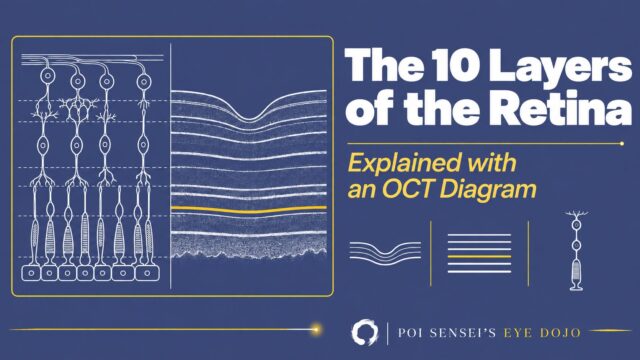
Once you can answer that, the classic lists stop being things to memorize. They become things you can work out at the viewing screen. FA also answers a different question from the OCT you already use every day: OCT shows you structure, FA shows you perfusion and leakage. Read the two together and most posterior segment findings start to explain themselves.
What FA actually adds
There are two dye studies, and they answer different questions.
- Fluorescein angiography (FA) shows you the retinal vessels.
- Indocyanine green angiography (ICGA) shows you the choroidal vessels.
FA is the one you will order most often. Its single greatest advantage over a color fundus photograph is that it tells you whether dye is escaping from vessels. That is why hyperfluorescence and hypofluorescence are the two findings worth reading carefully.
Start with one question, not a list
Textbooks give four causes of hyperfluorescence. The list is correct, but it is not how you read an angiogram. There is a simpler split underneath it:
- Dye is somewhere it should not be. It has escaped from vessels, collected in a space, or soaked into tissue that holds it.
- The filter in front of the choroid is gone. No dye is out of place. You are seeing the normal choroidal glow more clearly than usual.
Three of the four patterns belong to the first group. Only one belongs to the second. Decide which group you are in, and you are most of the way to the diagnosis.
The second habit that separates the patterns is time. An angiogram is a sequence, not a single picture, and each pattern behaves differently between the early and late phases:
| Pattern | Across the phases |
|---|---|
| Leakage | Brighter, larger, blurrier |
| Staining | Brighter, but same size and same border |
| Pooling | Progressively fills a defined space |
| Window defect | Bright early, fixed borders, does not grow |
Hold on to those two axes — where is the dye, and what does it do over time — and the rest of this article is just filling in examples.
Step 1: Decide whether the area is too bright or too dark
Compare the area with the surrounding retina, not with your memory of a normal study.
- Hyperfluorescence — brighter than it should be.
- Hypofluorescence — darker than it should be.
Do this before naming anything.
Step 2: If it is too bright, name the pattern
Leakage
Dye escapes from damaged vessels into the surrounding tissue. The bright area has a fuzzy border and spreads as the study goes on.
Macular edema is the everyday example. In diabetic macular edema and retinal vein occlusion, the inner blood-retinal barrier breaks down and dye leaks into the cystoid spaces around the fovea. In the late phase the spaces light up in the classic petaloid pattern.
In Behçet disease, the capillaries break down and fluorescein leaks out along the vessels themselves. The retinal vascular tree lights up in a branching pattern that is described as fern-like leakage.
Pooling
Dye collects in an anatomic space that can hold it — most often the subretinal or sub-RPE space.
In central serous chorioretinopathy, dye passes through a defect in the RPE and accumulates in the serous retinal detachment. In Vogt-Koyanagi-Harada disease, multiple leaking points fill the neurosensory detachments in the late phase. A pigment epithelial detachment behaves the same way: a defined space that grows steadily brighter while its border stays sharp.
Staining
Dye is taken up by tissue and stays there, so the structure itself glows. The area gets brighter in the late phase, but unlike leakage it keeps its size and its border.
In retinal vein occlusion you see leakage from the vessels in the occluded territory, but you also see the vein walls themselves shining. Increased permeability lets dye into the wall, and the wall retains it.
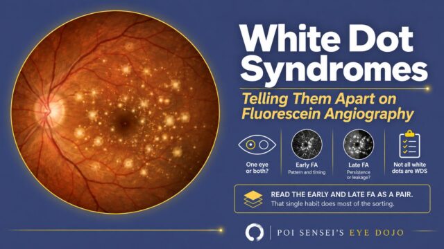
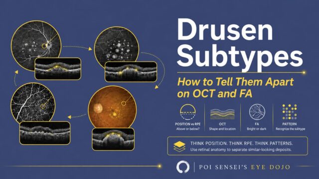
Staining does not always mean damage. Some structures simply hold dye: the normal optic disc, the sclera where it is exposed, fibrotic scars, and drusen all stain in the late phase without any vessel being broken. That is worth remembering before you call every late bright spot pathological. Drusen are not a single finding, though: whether a deposit looks bright or dark here depends on which side of the RPE it sits on, which is worked through in drusen subtypes on OCT and FA.
Window defect
This one is different. Nothing has leaked.
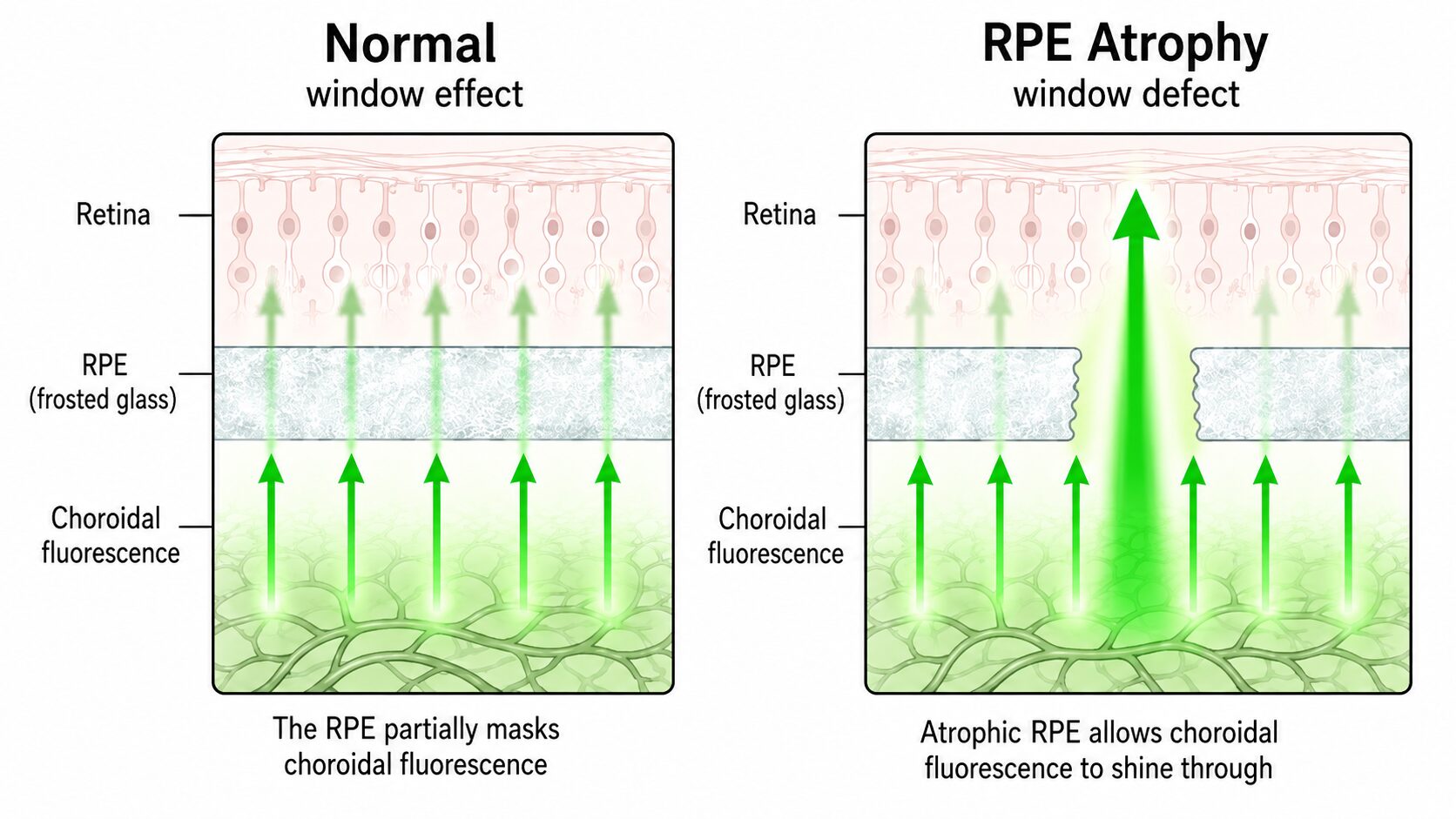
Look at a normal angiogram first. Dye enters both the retinal and the choroidal circulation. The retinal pigment epithelium (RPE) sits between the two, and it partly screens the choroidal fluorescence behind it. So the background of a normal angiogram glows faintly and evenly. That is the window effect — like looking at a lamp through frosted glass.
Now take the frosted glass away. In geographic atrophy the RPE is lost, so the choroidal fluorescence comes through far brighter in those areas. That is a window defect: a hole in the frosted glass, with the light shining straight through. It is bright from the earliest frames, its borders match the atrophy exactly, and it never grows.
Retinitis pigmentosa shows you both sides of the RPE at once: the atrophic areas transmit the choroidal glow and look bright, while the bone-spicule pigment clumps themselves block fluorescence and look dark. Keep the two apart — the atrophy is the window, the pigment is a blocker.
Step 3: Turn the pattern into anatomy
| What you see | What is wrong | Where |
|---|---|---|
| Leakage | Dye escaping through a broken blood-retinal barrier | Retinal vessels |
| Pooling | Dye collecting in a space that should not be there | Subretinal / sub-RPE space |
| Staining | Tissue holding on to dye | Vessel walls, scar, disc, sclera |
| Window defect | Loss of the screen in front of the choroid | RPE |
This is why the four-item list is worth reorganizing. The first three are all about dye out of place — where it escapes, where it collects, what holds it. Only the fourth is bright without any dye out of place.
Step 4: If it is too dark, decide why the light is missing
Hypofluorescence also has two mechanisms, and they are easy to separate.
The dye never arrived — a filling delay or filling defect. This means a circulatory problem in the retina or choroid. An incomplete retinal artery occlusion slows the dye down, so the territory fills late. A complete occlusion means it does not fill at all. In diabetic retinopathy, capillary closure produces dark territory called a non-perfusion area (NPA).
The dye arrived, but something in front is hiding it — blocked fluorescence. Anything sitting in front of the source of the fluorescence can block it: vitreous hemorrhage or opacity, preretinal or subretinal hemorrhage, and pigment or lipid deposits within the retina. Subretinal drusenoid deposits work the same way: they sit in front of the choroidal glow and block it, which is why they are dark here while ordinary drusen are bright. Depth decides how much disappears — subretinal blood hides the choroidal glow but leaves the retinal vessels visible, while a preretinal hemorrhage hides both.
The fundus photograph usually tells you which of the two you are looking at, because blockage has something visible doing the blocking.
Two traps
A dark macula is normal. On a healthy angiogram the retina around the macula glows faintly from the choroid behind it, while the macula itself stays dark. That contrast is the normal state, not hypofluorescence. Judge every area against the retina next to it, and remember what the center is supposed to look like.
A bright area that does not spread is not leakage. Leakage escapes into tissue, so its border blurs and the area grows as the study goes on. Staining and a window defect both stay inside their own borders from beginning to end. Before you call something leakage, watch it across the phases of the study rather than judging a single frame.
Neither of those has to be memorized. Both come from the same habit: read the study as a sequence, and compare each area with the retina beside it.
In short
- Ask two questions about every abnormal area: where is the dye, and what does it do over time?
- Leakage grows and blurs. Staining brightens without growing. Pooling fills a defined space.
- A window defect means the RPE is gone and the normal choroidal fluorescence is showing through — bright early, fixed borders, no growth.
- Dark areas are either never filled (circulation) or blocked (something in front of the light).
- When a finding surprises you, go back to the layer. The layer explains the picture. And when the question is function rather than structure or circulation, that is a different study — ERG.