PCV vs Type 3 MNV (RAP) vs Typical AMD: How to Tell Them Apart on FA and ICGA
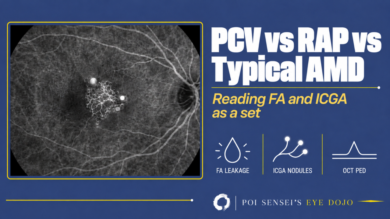
Neovascular age-related macular degeneration (AMD) is now classified by the type of macular neovascularization (MNV). In practice, three pictures are worth learning: type 1 or type 2 MNV without polypoidal lesions (often called typical neovascular AMD), polypoidal choroidal vasculopathy (PCV) and type 3 MNV, once called retinal angiomatous proliferation (RAP). Learn these three and you have the outline of neovascular AMD. They look different on the angiogram and respond differently to treatment. Read fluorescein angiography (FA) and indocyanine green angiography (ICGA) as a set, and check optical coherence tomography (OCT) with them.
- On FA, is the leak classic, with a clear outline from the early phase, or occult, with no clear leaking point?
- On ICGA, is the bright lesion a wide late plaque, early bright nodules or a small hot spot?
- Does a retinal vessel run into it?
- On OCT, how is the retinal pigment epithelium (RPE) raised?
Where the new vessels grow
New vessels in AMD are now called MNV. The old term, choroidal neovascularization (CNV), did not fit vessels that start in the retina. MNV is grouped by the layer it grows into.
- Type 1 MNV grows from the choroid through Bruch membrane and stays under the RPE.
- Type 2 MNV breaks through the RPE into the space under the retina.
- Type 3 MNV starts in the retina itself and grows down toward the RPE.
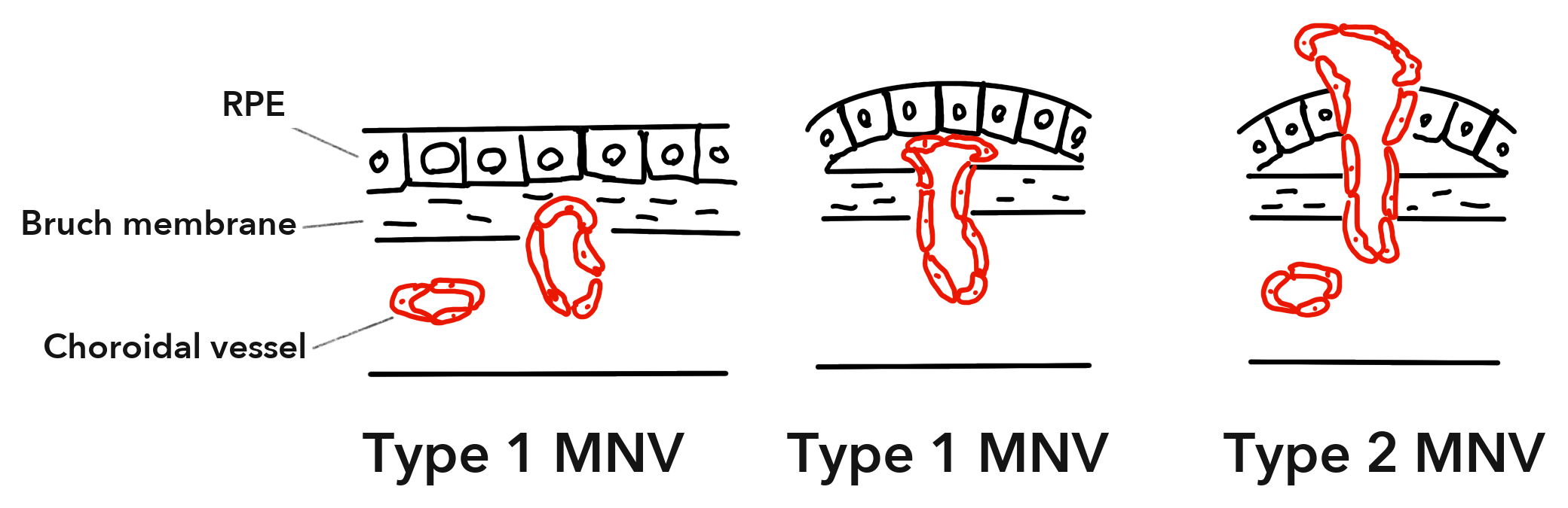
When type 1 and type 2 are both present, it is called mixed type 1 and type 2 MNV. PCV is type 1 MNV with polypoidal lesions at its edge.
FA and ICGA: what each one shows
FA and ICGA see different things, so use them together.
- FA uses fluorescein, a small molecule that leaks out of new vessels. It shows where MNV is and how much it leaks. But the RPE blocks much of its light, so vessels under the RPE are hard to see.
- ICGA uses indocyanine green, which binds to proteins in the blood and leaks much less than fluorescein. Its near-infrared light passes through the RPE, pigment and thin layers of blood, so ICGA shows the vessels under the RPE. It is the key test for telling PCV and type 3 MNV from type 1 or type 2 MNV.
On FA, leakage from MNV comes in two patterns.
- Classic leakage is a bright area with a clear outline in the early phase. It then leaks into the space under the retina, and its edges blur by the late phase. Put simply, you can see where the leak comes from.
- Occult leakage has no clear leaking point. The area over the raised RPE glows in diffuse, speckled dots that leak slowly.
Classic leakage usually comes from type 2 MNV, and occult leakage from type 1 MNV. The match is not exact.
Type 1 and type 2 MNV: a plaque
In type 1 or type 2 MNV without polypoidal lesions, drusen are usually present in one or both eyes.
On FA, the lesion shows classic leakage, occult leakage or both. Occult leakage is the more common.
On ICGA, MNV shows as a plaque or a hot spot. A plaque is a bright area larger than one disc diameter. It shows best in the late phase and leaks little. A hot spot is a bright spot smaller than one disc diameter. Plaques are more common, and most come from type 1 MNV.
A hot spot has three possible causes: PCV, type 3 MNV or a small type 1 MNV. So a hot spot is your cue to look for PCV and type 3 MNV.
On OCT, type 1 MNV lifts the RPE in a low, irregular bump. You see two lines, the RPE and Bruch membrane, with material between them. This double-layer sign points to type 1 MNV. Type 2 MNV shows as bright (hyperreflective) material above the RPE, under the retina.
PCV: bright nodules in the early phase
In PCV, type 1 MNV spreads as a branching neovascular network (once called the branching vascular network) and ends in round, aneurysm-like dilated vessels called polypoidal lesions. The polypoidal lesions leak and bleed. The fluid or blood lifts the RPE off Bruch membrane. This is a pigment epithelial detachment (PED), and in PCV it happens again and again.
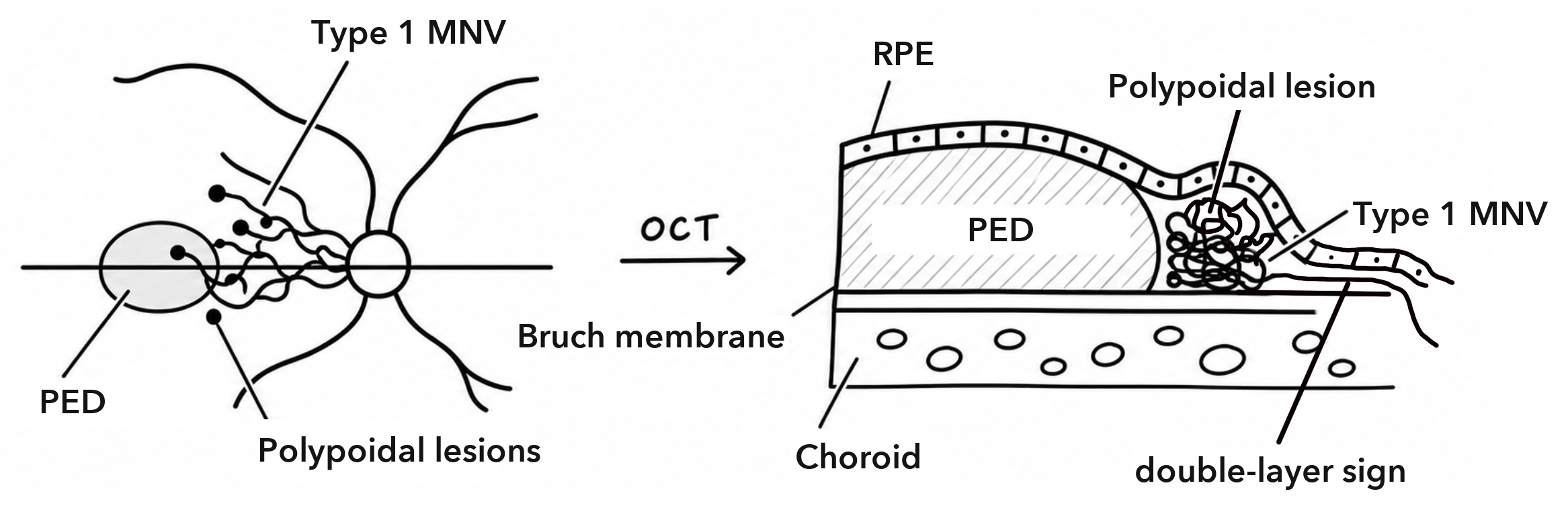
On the fundus, polypoidal lesions may be seen as orange-red nodules under the RPE, near the disc or in the macula. PCV can bleed heavily. A large subretinal hemorrhage should make you think of PCV or a ruptured retinal arterial macroaneurysm, and ICGA helps tell them apart. Drusen are often absent. If the fellow eye has no soft drusen, PCV is more likely than type 1 or type 2 MNV with drusen.
On FA, PCV usually shows occult leakage. A polypoidal lesion may show as a small, intensely bright spot, and dye slowly pools in the fluid of a PED. The polypoidal lesions and the type 1 MNV themselves are hard to see on FA.
ICGA is the key test. In the early phase, bright nodules appear at the edge of the type 1 MNV, often with a dark halo around them. The polypoidal lesions soon leak, and the area around them turns bright by the late phase. Late on, a polypoidal lesion can also look like a bright ring with a dark center. In clinical trials, PCV is diagnosed by a focal bright spot within the first 6 minutes of ICGA, plus one more feature such as nodules, a dark halo or the network of type 1 MNV[1].
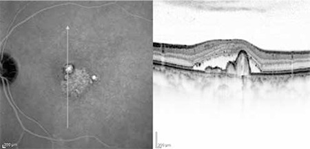
On OCT, a polypoidal lesion lifts the RPE steeply into a sharp-peaked PED. Under the raised RPE, the polypoidal lesion often shows as a round structure with a bright outline and a dark center, a sub-RPE ring-like lesion. Next to it, the type 1 MNV gives a flat, irregular PED with a double-layer sign.
Without ICGA, OCT can still point to PCV. A 2021 Asia-Pacific consensus uses three OCT signs: a sharp-peaked PED, a sub-RPE ring-like lesion and, on en face OCT, a complex RPE elevation where the branching network joins several PEDs. When all three are present, PCV is very likely, with a specificity of 91%. Their absence does not rule PCV out, because the sensitivity is 75%[2].
PCV is far more common in Asian patients. Among patients with neovascular AMD, it accounts for about 4% to 14% in White patients and 22% to 62% in Asian patients[1].
PCV is treated with anti-VEGF injections, alone or with photodynamic therapy (PDT). In the EVEREST II trial, adding PDT to ranibizumab closed more polypoidal lesions and needed fewer injections. If neovascular AMD has not responded after 3 months of anti-VEGF injections, suspect PCV and check with ICGA.
Type 3 MNV (RAP): a hot spot fed by a retinal vessel
In type 3 MNV, new vessels start in the deep capillary plexus of the retina and grow down through the outer retina. Later they can break through the RPE and join vessels from the choroid, a retinal-choroidal anastomosis.
Type 3 MNV affects older patients, more often women. It accounts for about 10% to 20% of neovascular AMD in White patients. It is often bilateral, so check the fellow eye for an early lesion. Eyes with type 3 MNV usually have many soft drusen and subretinal drusenoid deposits (SDD). SDD are a strong risk factor for type 3 MNV. The choroid is often thin.
On the fundus, small intraretinal hemorrhages near the fovea, in an eye full of soft drusen, suggest type 3 MNV.
On FA, type 3 MNV leaks into the retina, often like cystoid macular edema. Speckled bright dots may also appear over a raised RPE and spread in later frames, much like occult leakage. The late phase leaks heavily.
ICGA shows the lesion more clearly and makes the diagnosis in most cases. It shows a small hot spot in the mid or late phase. In the early phase, a retinal arteriole may run down into the lesion and a venule may drain it, sometimes joined as a hairpin loop. If you can see that link with a retinal vessel on FA or ICGA, the diagnosis is almost certain.
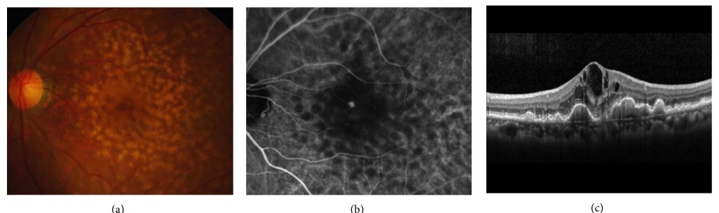
On OCT, intraretinal fluid shows early, often like cystoid macular edema. A PED follows as the disease goes on. Where the new vessels reach the PED, they show as a small bright lesion just above the RPE. The RPE line under that lesion is broken. This is the bump sign, and it strongly suggests type 3 MNV.
Type 3 MNV responds quickly to anti-VEGF injections, especially early, and the fluid usually clears. But it tends to come back, so injections must continue. Over the years, many eyes develop macular atrophy, which limits vision. One study found atrophy more often after aflibercept than after ranibizumab (43% vs 19% at 1 year), perhaps because aflibercept thins the choroid more and the choroid in type 3 MNV is already thin[3]. A longer study found no clear difference[4]. PDT alone works poorly, and Japanese guidelines no longer recommend PDT for type 3 MNV, because it can make atrophy worse.
Summary
Read FA and ICGA as a set: FA shows the leak, and ICGA shows the vessels under the RPE.
| Type 1 or 2 MNV | PCV | Type 3 MNV (RAP) | |
|---|---|---|---|
| FA | Classic, occult or both | Occult; polypoidal lesions hard to see | Leak into the retina; heavy late leak |
| ICGA | Late plaque; can be a small hot spot | Early bright nodules at the edge of type 1 MNV | Mid- or late-phase hot spot fed by a retinal vessel |
| OCT | Type 1: low RPE lift with a double-layer sign. Type 2: bright material above the RPE | Sharp-peaked PED and a sub-RPE ring-like lesion, with a double-layer sign next to it | Fluid in the retina; bump sign on a PED |
A hot spot on ICGA can be PCV, type 3 MNV or a small type 1 MNV.
References
[1] Chaikitmongkol V, Cheung CMG, Koizumi H, Govindahar V, Chhablani J, Lai TYY. Latest developments in polypoidal choroidal vasculopathy: epidemiology, etiology, diagnosis, and treatment. Asia Pac J Ophthalmol (Phila). 2020;9(3):260-268. PMID 32332215.
[2] Cheung CMG, Lai TYY, Teo K, et al. Polypoidal choroidal vasculopathy: consensus nomenclature and non-indocyanine green angiograph diagnostic criteria from the Asia-Pacific Ocular Imaging Society PCV Workgroup. Ophthalmology. 2021;128(3):443-452. PMID 32795496.
[3] Cho HJ, Hwang HJ, Kim HS, Han JI, Lee DW, Kim JW. Intravitreal aflibercept and ranibizumab injections for type 3 neovascularization. Retina. 2018;38(11):2150-2158. PMID 28984737.
[4] Kim JH, Kim JW, Kim CG, Lee DW. Long-term treatment outcomes in type 3 neovascularization: focus on the difference in outcomes between geographic atrophy and fibrotic scarring. J Clin Med. 2020;9(4):1145. PMID 32316276.

Which retinal layer makes which wave, the three abnormal patterns, and the dark-adaptation trap to check before you report “negative” — on one page, by email. One email when I publish something new, about once a month. Unsubscribe anytime.