How to Read a Macular OCT: A Step-by-Step Approach
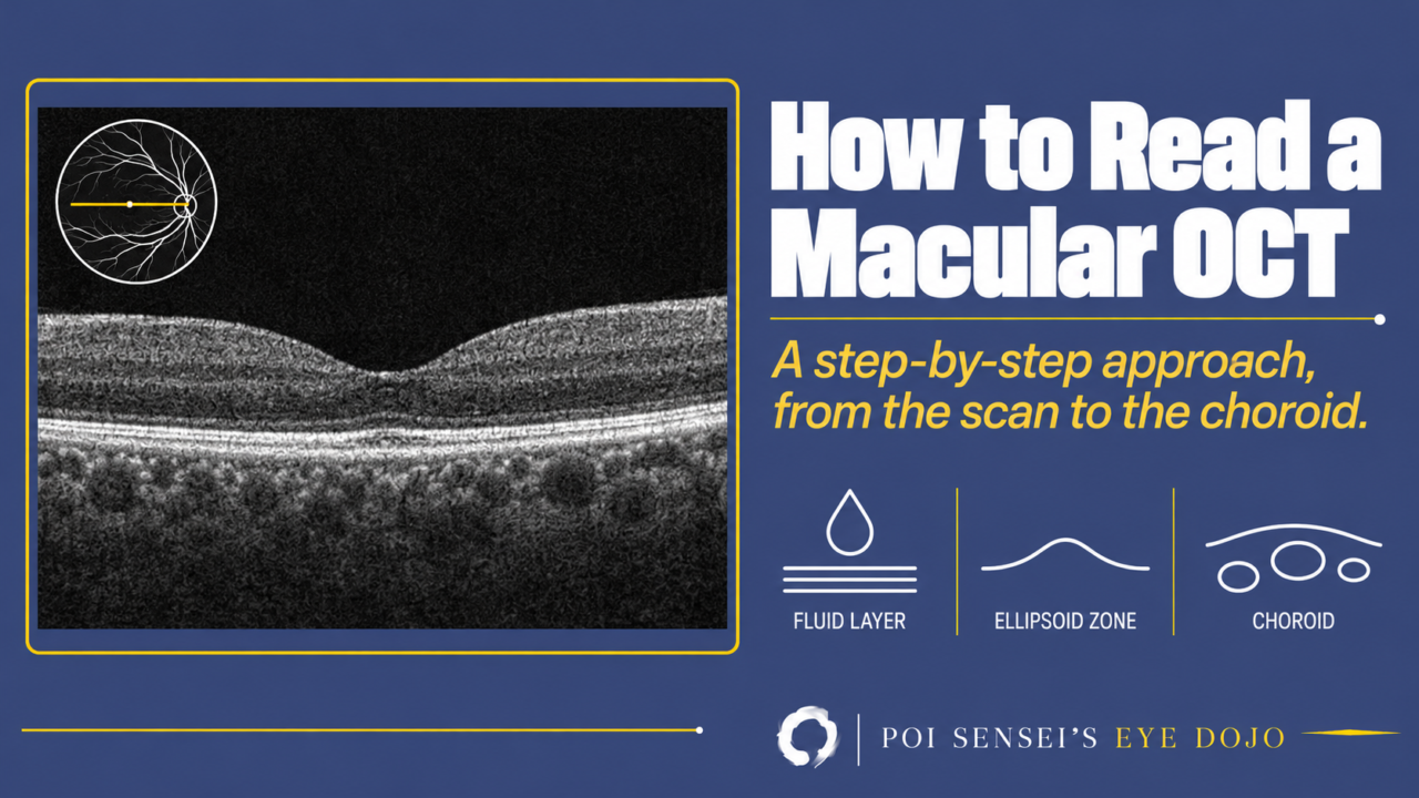
Read a macular OCT in the same order every time, from the scan itself to the choroid. When you find fluid, name the layer it is in before you name the disease.
- Check the eye, the scan position and the image quality.
- Find any fluid and name the layer it is in.
- Look for deposits.
- Check the retinal pigment epithelium (RPE).
- Check the ellipsoid zone (EZ) and the foveal bulge.
- Look at the thickness of the choroid.
The normal macula on OCT
At the center of the fovea, the inner retina is pushed aside, so light reaches the photoreceptors directly. Before you read an abnormal scan, find each layer on a normal one. For the cells in each layer, see the 10 layers of the retina.
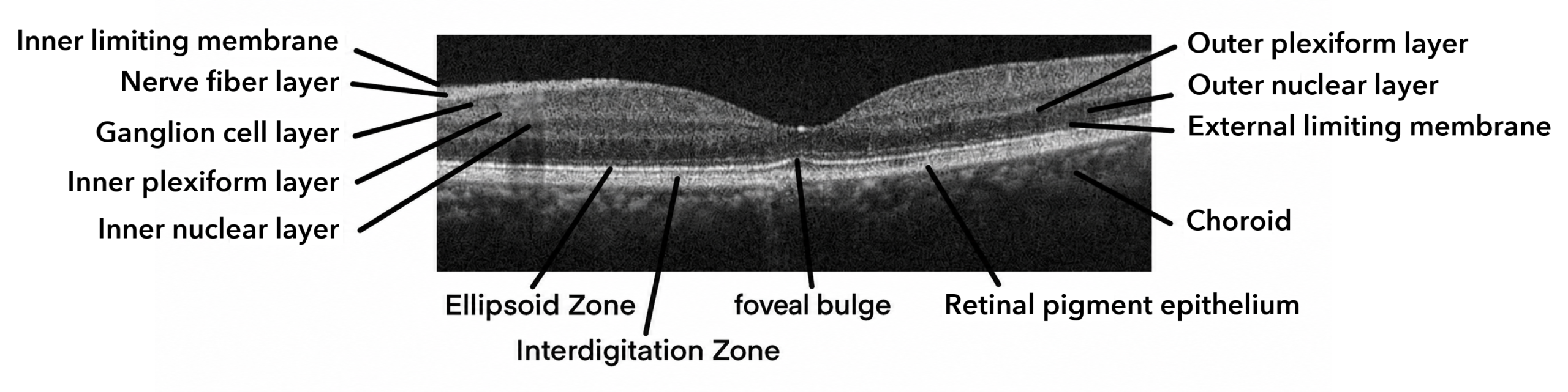
Before you read: three checks
Check three things first: which eye it is, whether the scan passes through the fovea, and whether the image is good enough (signal strength, fixation, artifacts). If you skip this, you will read a scan that misses the fovea as a loss of the foveal pit.
Then describe the finding before you name a disease. Say which layer the fluid or the elevation is in: intraretinal cysts, subretinal fluid, or an elevation under the RPE. Once you can describe it, you can discuss the case with a senior colleague and search for the finding. My tip is an image search with those words. You will see scans that look like your patient’s.
Step 1: Which layer is the fluid in?
In age-related macular degeneration (AMD) and diabetic macular edema, you will usually see at once that something is wrong. The next step is to name the layer.
| Where the fluid is | Think of first |
|---|---|
| Inside the retina, in the inner nuclear and outer plexiform layers | Diabetic retinopathy, retinal vein occlusion |
| Under the retina, above an abnormal RPE | AMD, central serous chorioretinopathy (CSC) |
In diabetic retinopathy and retinal vein occlusion, the inner blood-retinal barrier (the walls of the retinal vessels) breaks down, and the retinal vessels leak into the retina. In CSC, the outer blood-retinal barrier (the RPE) breaks down, and fluid from the choroid leaks under the retina. Also look for thinning of a single layer, such as the ganglion cell layer or the outer nuclear layer.
Step 2: Look for deposits
When a white lesion on the fundus puzzles you, put an OCT scan through it. Hard exudates usually sit in the outer plexiform layer, between the inner and outer retina. Drusen usually sit under the RPE.
Step 3: Check the RPE
In AMD, new vessels grow from the choroid (macular neovascularization). They can lift the RPE, which is called a pigment epithelial detachment (PED). They can also break through the RPE and grow toward the retina. A wavy RPE across the whole scan is seen in hypotony (very low eye pressure) and in Vogt-Koyanagi-Harada (VKH) disease.
Step 4: Check the EZ and the foveal bulge
Of the bright bands in the photoreceptor layer, the ellipsoid zone (EZ) is thought to match the ellipsoid of the inner segments, the part packed with mitochondria. It used to be called the junction of the inner and outer segments (IS/OS line). The EZ is very useful for judging whether the photoreceptors are damaged.
- In retinitis pigmentosa, the EZ fades where the visual field is lost.
- In occult macular dystrophy, the fundus looks normal, and OCT shows a blurred EZ at the fovea.
- In acute zonal occult outer retinopathy (AZOOR) and cancer-associated retinopathy, EZ loss that matches the field defect helps the diagnosis.
When loss of acuity or field has no clear cause, look at the EZ. A blurred EZ is easy to miss until you are used to it. Do not look at the scan vaguely; follow the EZ on purpose, every time.
The cones at the fovea are tall, like rods, so the EZ rises slightly there. This is the foveal bulge. In some patients whose vision does not recover after treatment for retinal vein occlusion or CSC, the EZ is preserved but the foveal bulge is gone.
Step 5: Look at the choroid
Modern OCT shows the choroid too. A choroid that is thick for the patient’s age and refraction, with large vessels, is called pachychoroid. CSC and polypoidal choroidal vasculopathy (PCV) develop on a pachychoroid. In acute VKH disease, the choroid thickens and then thins with treatment, so its thickness shows the response.
Summary
- Check the eye, the scan through the fovea and the image quality first.
- Name the layer the fluid is in before you name the disease.
- Then check deposits, the RPE, the EZ and the foveal bulge.
- Finish with the thickness of the choroid.

Which retinal layer makes which wave, the three abnormal patterns, and the dark-adaptation trap to check before you report “negative” — on one page, by email. One email when I publish something new, about once a month. Unsubscribe anytime.