Color Vision Tests: Ishihara, D-15 and the Anomaloscope
A positive Ishihara test tells you that a patient probably has a red-green color vision defect. It does not tell you how strong the defect is or exactly which type it is. That takes two more tests: the D-15 and the anomaloscope. This article shows what each test answers and how to read the results.
- Screen with pseudoisochromatic plates such as the Ishihara.
- Grade the severity with the D-15.
- Confirm the type with the anomaloscope.
How color vision works
The retina has three types of cones. Each type responds best to a different wavelength: S cones (short, blue) near 420 nm, M cones (middle, green) near 530 nm and L cones (long, red) near 560 nm.
We see color by comparing how strongly the three types respond. When all three respond fully, we see white. When none respond, we see black.
Types of color vision deficiency
A color vision deficiency is named after the cone type that is affected. A problem with the L cones is called protan, with the M cones deutan, and with the S cones tritan. Protan and deutan defects are red-green defects; tritan defects are blue-yellow.
Anomalous trichromacy, dichromacy and monochromacy
In anomalous trichromacy, all three cone types are present, but one of them works abnormally. An abnormal L cone causes protanomaly, and an abnormal M cone causes deuteranomaly.
In dichromacy, one cone type is missing. A missing L cone causes protanopia, and a missing M cone causes deuteranopia.
In monochromacy, only one type of photoreceptor works for color. In achromatopsia (rod monochromacy), no cones work. In blue cone monochromacy, only the S cones work. Either way, the patient cannot tell colors apart, because the brain sees color by comparing the signals from two or more cone types. Visual acuity is reduced in both, but it is often a little better in blue cone monochromacy, because some cones still work.
Dichromacy is often more severe than anomalous trichromacy. But the severity varies from person to person, and the symptoms are very similar. So protanomaly and protanopia are often grouped together as a protan defect, and the same goes for deutan defects.
Inheritance and how common it is
Protan and deutan defects are X-linked recessive, so they are much more common in men. They affect about 8% of men of Northern European descent and 0.5% of women. In Japan, the numbers are about 5% of men and 0.2% of women. Deutan defects are about three times as common as protan defects.
Inherited tritan defects are rare and autosomal dominant. Most tritan defects are acquired, from aging or retinal disease such as central serous chorioretinopathy. In general, macular disease tends to cause blue-yellow defects, and optic nerve disease tends to cause red-green defects. Blue cone monochromacy is X-linked recessive.
What patients actually see
A protan defect does not mean the patient cannot see red. Patients with a red-green defect still see red as red and green as green. What changes is that the boundaries between colors become less clear. As a rough picture (the numbers are only an illustration, not measured values), a color gradient that a normal eye sees in 10 steps may look like only 7 steps to a person with a red-green defect. In protan defects, reds also look darker.
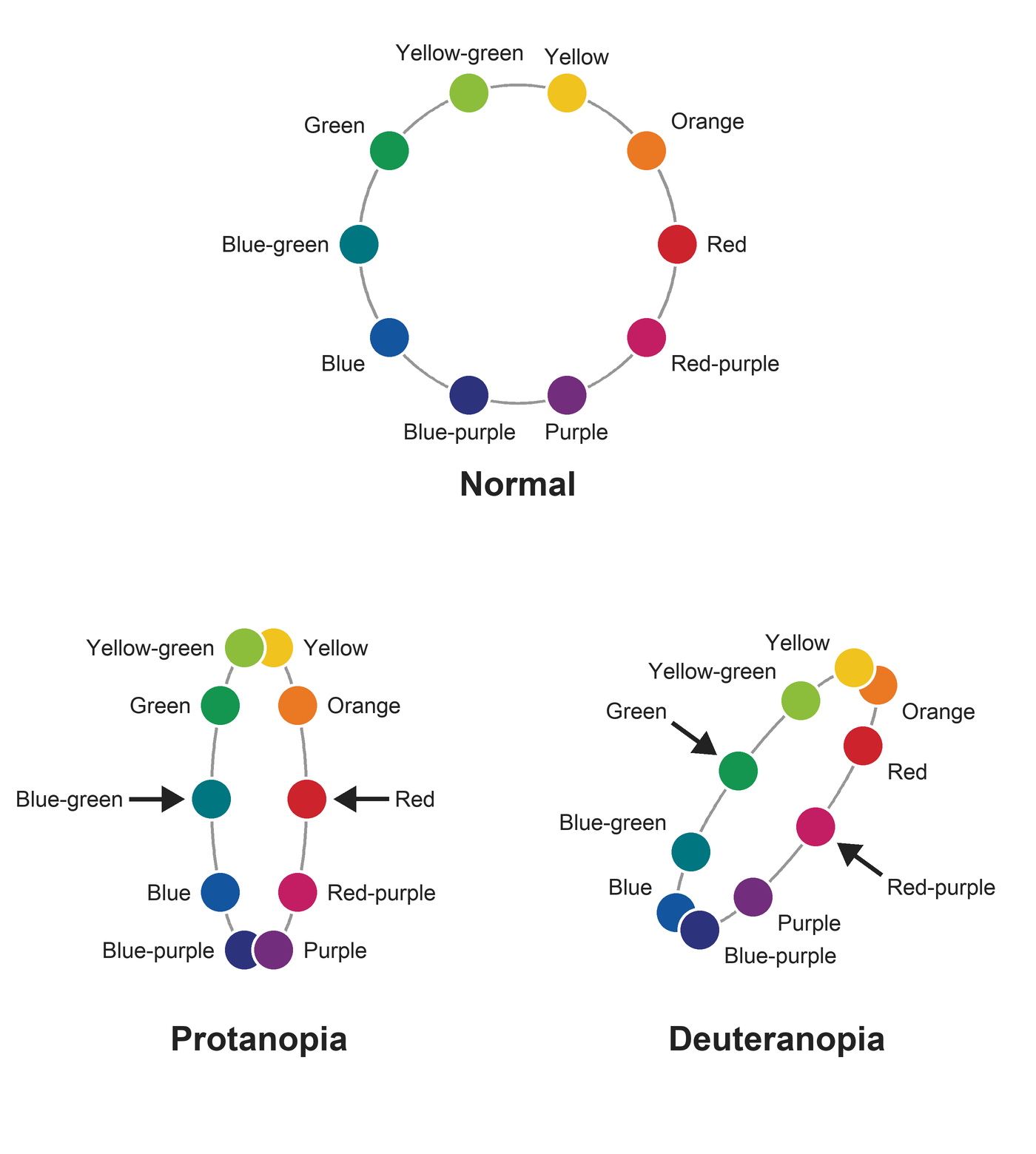
In this diagram, 10 hues sit on a circle, and colors facing each other are complementary. In a red-green defect, the circle is squeezed into a thin ellipse. In protanopia, it is squeezed from the sides, so red comes close to blue-green and green comes close to orange. In deuteranopia, it is squeezed at a slant, so green comes close to red-purple. Colors that end up close are hard to tell apart. Blue and yellow stay far apart, so they can be told apart without trouble. And as the figure shows, warm and cool colors stay relatively far apart, so a warm color next to a cool color is less likely to be hard to see. In anomalous trichromacy, the circle is squeezed less.
- Red and green (red strawberries among green leaves)
- Red and black (red and black ballpoint pens)
- Blue and purple
- Pink and gray
- Light blue and pink
Step 1: Screen with pseudoisochromatic plates
Pseudoisochromatic plates show a number made of colored dots on a background of other dots. A person with normal color vision and a person with a red-green defect see different things. For example, a normal eye reads one plate as 8, while an eye with a red-green defect reads it as 3. The Ishihara test is the best-known example.
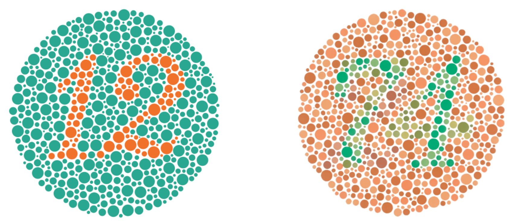
Everyone can read the plate on the left (12), whatever their color vision. The plate on the right reads 74 with normal color vision, but it is hard to read with a red-green defect. I have protanopia myself, and I cannot read the 74. Colors on a screen differ from the printed plates, so do not use this image to test anyone.
The Ishihara test is very sensitive for red-green defects, so it is used for screening. On the other hand, it cannot grade the severity or tell protan from deutan. It also misses tritan defects. If you suspect a tritan defect, use the Hardy-Rand-Rittler (HRR) plates, which detect all three types and grade protan and deutan defects as mild, medium or strong.
The 12 plate is the first plate of the test. If a patient with good visual acuity cannot read it, suspect non-organic vision loss.
For a suspected congenital defect, test with both eyes open. For a suspected acquired defect, test each eye separately, because the disease may affect only one eye.
Step 2: Grade the severity with the D-15
The Farnsworth D-15 has 15 colored caps and one fixed reference cap. The patient places the caps in order, each next to the one that looks most similar in color. You then connect the cap numbers on the score sheet in the order the patient placed them.
The figures below are D-15 score sheets. Start with the pass pattern, and compare the others with it.
Pass with minor errors
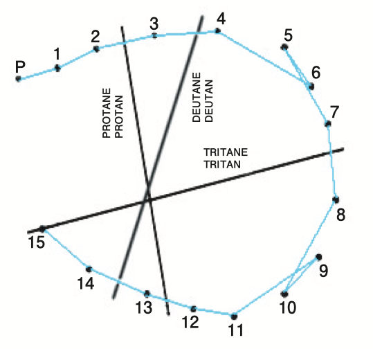
The line runs around the circle from 1 to 15. Here caps 5 and 6, and caps 9 and 10, are swapped. These swaps between neighbors are minor errors, and the result is a pass.
One crossing line
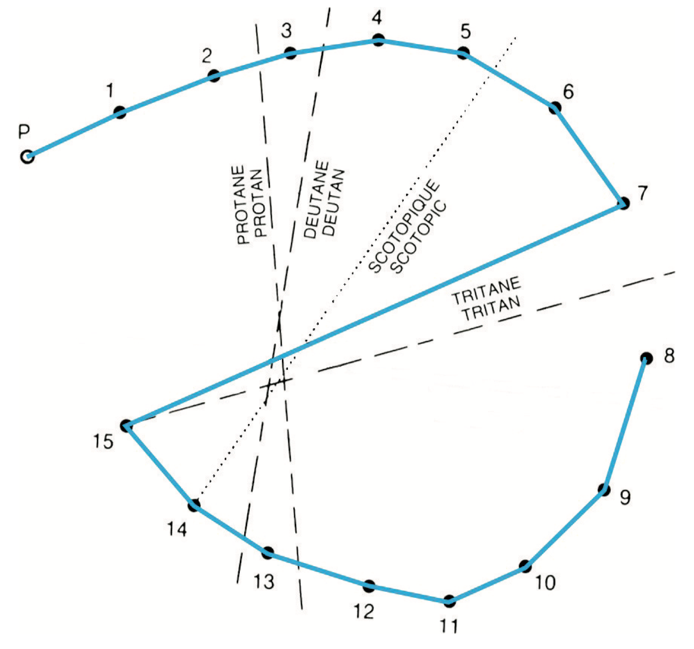
Here the patient placed caps 1 to 7 in order, jumped to 15, and then placed the rest from 14 back down to 8. This makes one line across the circle. This pattern is often seen even in people with normal color vision. Repeat the test. If the result is the same, count it as a pass.
Fail: two or more crossing lines
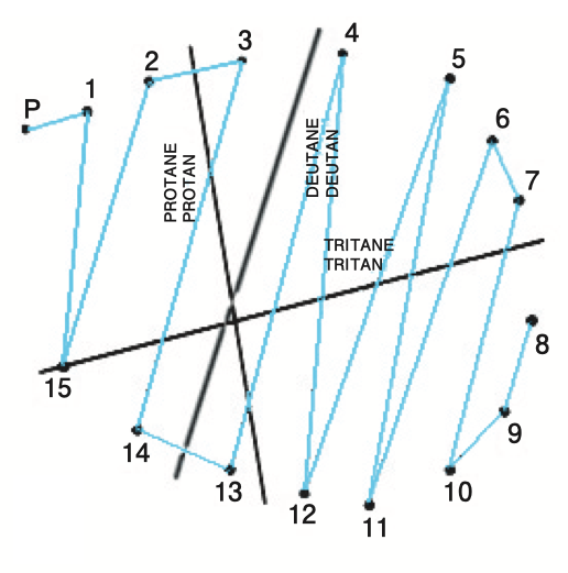
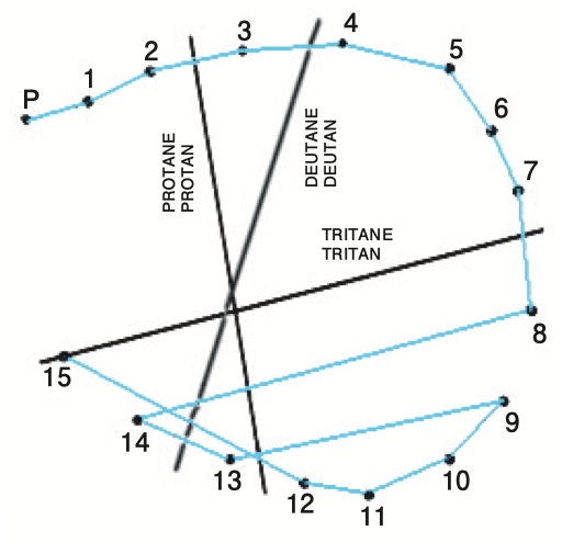
When two or more long lines cut across the circle, the result is a fail. The direction of the lines shows the type. The score sheet has three printed axes, labeled protan, deutan and tritan. Look for the axis the crossing lines run parallel to.
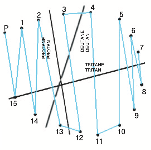
These crossing lines run parallel to the protan axis. This is a protan defect.
These lines run parallel to the deutan axis.
Here the lines from 8 to 14 and from 9 to 13 run parallel to the tritan axis. This is a tritan defect.
If the crossing lines do not run parallel to any axis, the result is a fail, but you cannot tell the type. Rod monochromacy gives its own pattern along a fourth axis, labeled scotopic on some score sheets.
What a pass means
The D-15 picks up only moderate and severe defects[1]. So a pass means the defect is mild or absent. A fail means the defect is moderate or severe.
Dichromats tend to fail and anomalous trichromats tend to pass, but there is overlap. I am a protanope, yet I pass the D-15.
Step 3: Confirm the type with the anomaloscope
The anomaloscope is the gold standard for diagnosing red-green defects. The patient looks into the instrument and sees a circle split into two halves. One half is a mixture of red and green light. The other half is pure yellow. The patient turns two knobs, one for the red-green mixture and one for the brightness of the yellow, until the two halves look the same. You change the mixture step by step and plot every setting the patient accepts as a match.
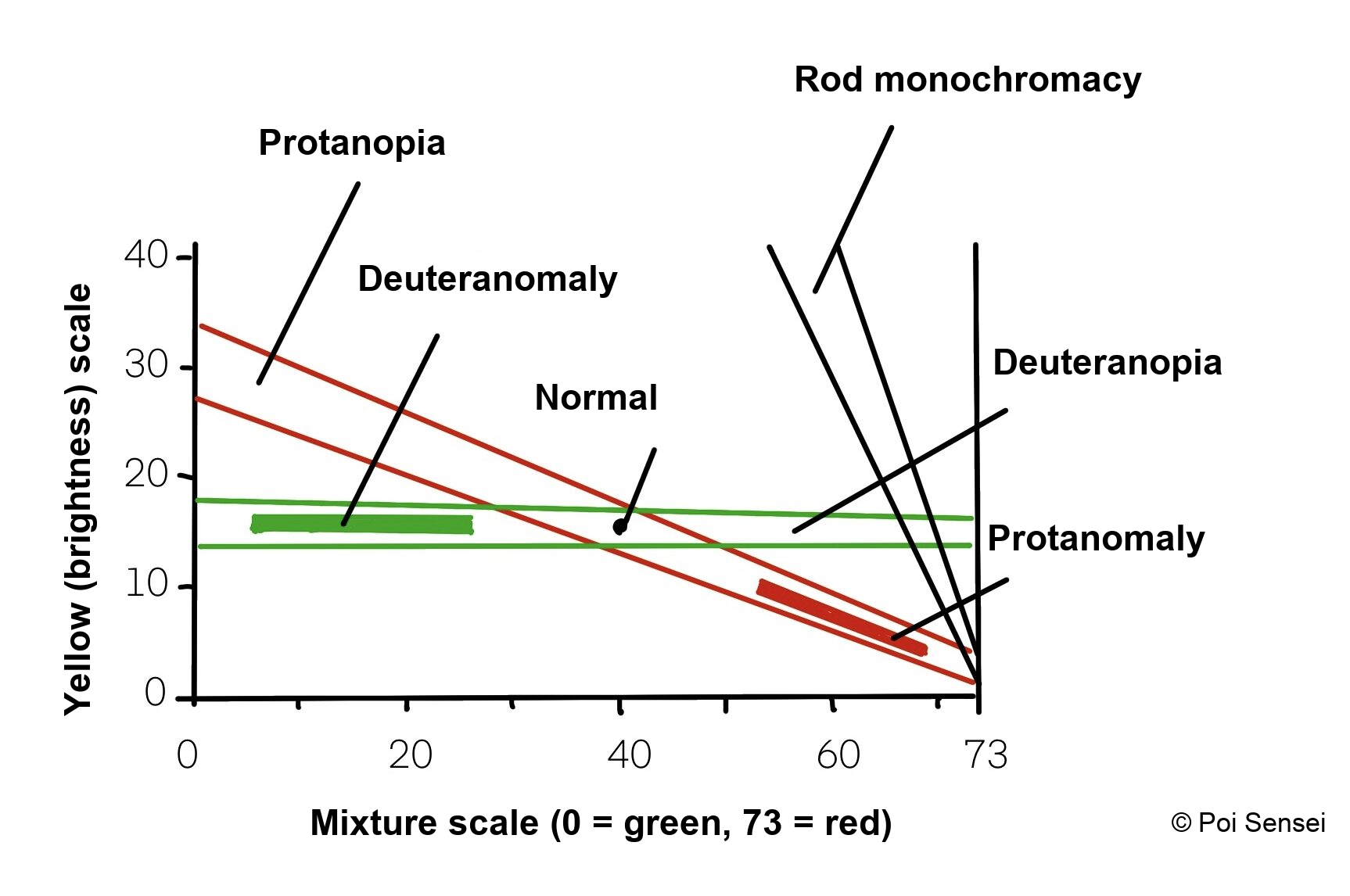
In this graph, the horizontal axis is the mixture scale (0 is pure green, 73 is pure red), and the vertical axis is the brightness of the yellow.
A normal eye matches in only a narrow range.
A dichromat accepts a match across the whole mixture scale, so the result is a line. In protanopia, the line falls toward the red end. Red looks dark, so the patient turns the yellow down to match it. In deuteranopia, the line is almost flat.
In anomalous trichromacy, the match range is shifted. In deuteranomaly, the patient needs more green, so the range is on the green side. In protanomaly, the patient needs more red, so the range is on the red side and the yellow is set darker.
In rod monochromacy, the line is very steep.
The anomaloscope is the test that separates dichromacy from anomalous trichromacy. Its red-green match does not test tritan defects.
It is hard to picture this test from a description. If your clinic has an anomaloscope, try it on yourself once. It makes much more sense after you have done it.
Color vision and jobs
Some jobs set color vision standards, such as pilots, ship crews, train drivers and the military. Jobs where color judgment matters, such as cooks who check the freshness of food, painters, artists, photographers and designers, can be harder. Still, many people with color vision deficiency do well in these fields. The standards differ by country and by employer.
When a child is diagnosed, the family usually worries most about the future. Knowing which jobs set standards helps you answer them.
The tests differ by country, too. After the Ishihara, some use a lantern test, which shows signal-colored lights for a short time and checks whether the person can name them quickly (for example, pilots in Australia and deck officers on US ships). Others use a computer test such as the CAD test. UK ship crews switched from the lantern to the CAD in 2018, and US pilots have taken a computer test since 2025. People with a color vision deficiency can often name the colors correctly when they look slowly, but make mistakes when they must answer fast. I can tell blue from purple when I look calmly, but in a fast color-matching puzzle game I often mix them up. I think this is exactly why jobs like pilot, where a split-second color judgment can cost lives, have color vision standards.
Summary
- The Ishihara plates screen for red-green defects but miss tritan defects.
- On the D-15, two or more crossing lines mean a fail, and their direction shows the type.
- Passing the D-15 means the defect is not severe, not that color vision is normal.
- The anomaloscope confirms the type and separates dichromacy from anomalous trichromacy.
References
[1] Costa MF, Henriques LD, Souza GS. An integrative review for clinical evaluation of color vision: the right test for the right disease. J Curr Ophthalmol. 2024;36(4):355-364. PMID 41041026.
Which retinal layer makes which wave, the three abnormal patterns, and the dark-adaptation trap to check before you report “negative” — on one page, by email. One email when I publish something new, about once a month. Unsubscribe anytime.