Hydroxychloroquine Retinopathy Screening: What to Look For on OCT and FAF
Hydroxychloroquine (Plaquenil) is a useful drug for lupus and rheumatoid arthritis. But after years of use it can damage the retina, and the patient usually notices nothing until the damage is advanced. Advanced damage can keep getting worse even after the drug is stopped. So the only way to protect vision is to find it early. This article shows how to screen: who is at risk, which tests to use and what the early signs look like.
- Check the dose and the risk factors.
- Get a baseline OCT and autofluorescence, then screen every year.
- Look in both the parafoveal and the pericentral area.
- Read the OCT and autofluorescence for early signs.
- Confirm unclear findings with a visual field or mfERG.
Why the retina is at risk
Hydroxychloroquine is used for systemic lupus erythematosus (SLE), cutaneous lupus and rheumatoid arthritis. In the eye, it is concentrated in tissues that contain melanin, such as the retinal pigment epithelium (RPE) and the choroid. The exact mechanism of damage is still unknown.
The damage starts in the photoreceptors and the outer retina. The RPE is affected later. Central visual acuity is kept until late, so most patients have no symptoms. A few notice small blind spots next to the center while reading.
Step 1: Check the dose and the risk factors
The recommended daily dose is 5.0 mg/kg of real body weight or less. Above this the risk rises, and near 6.0 mg/kg it rises sharply. In severe obesity, a dose based on weight can be too high, so the maximum is 400 mg a day.
The risk also grows with the years of use. At 5.0 mg/kg or less, the risk is below 1% within 5 years and below 2% at 10 years[1].
These factors raise the risk:
- A daily dose above 5.0 mg/kg
- Use for more than 5 years
- Kidney disease
- Tamoxifen use
- Starting the drug at an older age
Macular disease makes the screening tests harder to read. Whether it also raises the risk is not proven.
Check the dose against the weight at every visit, and ask about kidney disease, tamoxifen and weight loss.
Step 2: Baseline, then every year
Get a baseline examination close to the start of treatment: fundus examination, OCT and wide-field fundus autofluorescence (FAF). It rules out a maculopathy that is already there, and it gives you images to compare with later.
Then screen every year. If there are no risk factors, the early risk is very low, so the yearly screening can wait until 5 years of use. If there are risk factors, start it from the beginning.
| Role | Tests |
|---|---|
| Main tests | OCT, wide-field FAF |
| To confirm | Visual field, mfERG |
| Not for screening | Fundus exam alone, color vision, Amsler grid, full-field ERG |
Color vision tests such as the Ishihara chart and the full-field ERG change only late. A normal fundus does not rule out toxicity.
Step 3: Look in both areas
The damage usually starts in one of two areas. Which one depends partly on the patient’s background, but screen both areas in every patient.
Parafoveal pattern
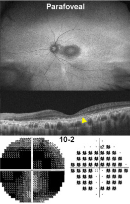
The damage forms a ring just outside the fovea. This is the usual pattern in patients of European descent, and also in those from India, Pakistan and the Middle East. The 10-2 visual field covers this area.
In this figure, the FAF is at the top, the OCT in the middle and the 10-2 visual field at the bottom. The arrowhead on the OCT points to the loss of the outer retina. This is an advanced case, so the ring is already dark on the FAF. Look at where the ring is, not how dark it is. Early on, the same area is thin on OCT and bright on FAF.
Pericentral pattern
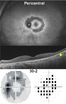
The damage starts farther out, near the vascular arcades. This is the usual pattern in East Asian patients. A standard macular OCT scan may not reach this area. You need wider OCT scans, wide-field FAF that extends beyond the arcades, and a 24-2 or 30-2 visual field.
The figure has the same layout. The dark ring sits outside the macula, and the arrowhead on the OCT is near the edge of the scan.
Step 4: Read the OCT and FAF
OCT: thinning comes first
The earliest sign is thinning of the outer retina. It shows on the thickness map, not on a single cross-section (B-scan). Compare the thickness values in each sector of the ETDRS grid with last year’s. Thinning can appear several years before any line on the B-scan changes.
Then the outer lines are lost in this order. If you are unsure where these lines are, see how to read a macular OCT.
- The interdigitation zone fades in the parafovea.
- The ellipsoid zone becomes thin or broken, then is lost.
- Loss of the external limiting membrane (ELM) marks severe damage.
The center is spared, so the preserved fovea stands out against the damaged ring. This is the "flying saucer" sign.
FAF: bright first, dark later
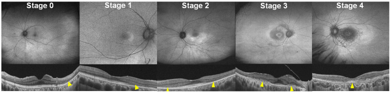
FAF is on the top row and OCT on the bottom. The arrowheads mark loss of the outer retina.
The first change is a bright area, most often in the inferotemporal macula (stage 1). It can grow into a partial or full ring (stage 2). The OCT shows thinning and ellipsoid zone loss in the same place.
Later, dark areas appear (stage 3). They mean the photoreceptors are gone and the RPE is damaged. In stage 4 the dark area reaches the fovea.
Compare with the baseline and last year’s images. A single image is not always easy to read. The goal of screening is to find toxicity before any area turns dark.
Step 5: Confirm with the visual field or mfERG
When the OCT or FAF is unclear, a functional test can confirm the finding.
Visual field
Use the 10-2 for the parafovea and the 24-2 or 30-2 for the pericentral area. Newer patterns such as the 24-2C cover both in one test. The earliest defects are often superonasal. The field is flipped compared with the retina, so this matches inferotemporal damage on the OCT and FAF. Repeat an uncertain field within a few months.
mfERG
The mfERG records the electrical response of many small areas of the retina, so it is an objective test. In the parafoveal pattern, the waves become small in the ring just around the fovea. In the pericentral pattern, they become small in the outer rings. Noise and artifacts can look like disease. See how to read a multifocal ERG.
A bull’s eye is a late sign
The classic bull’s eye maculopathy is a ring of RPE atrophy around a spared fovea. In the pericentral pattern, it appears near or beyond the arcades. When you can see it on the fundus, the RPE is already damaged. With proper dosing and yearly screening, it should no longer be seen.
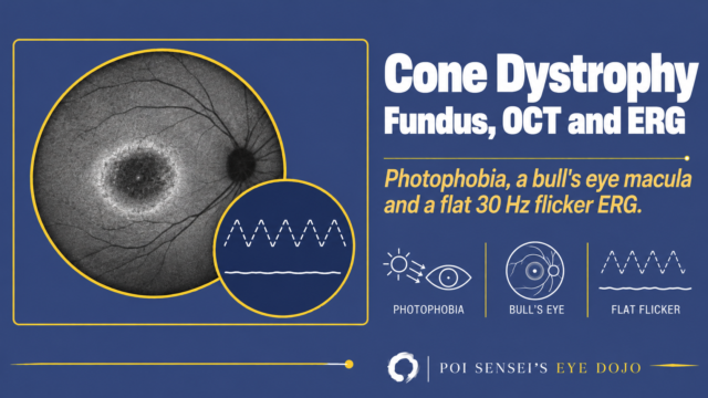
A bull’s eye can also come from other diseases, such as cone dystrophy and Stargardt disease. Check the drug history. End-stage toxicity can even look like retinitis pigmentosa, with widespread atrophy and a reduced full-field ERG.
If you find toxicity
Whether to stop the drug is decided with the patient and the prescribing physician, not by the ophthalmologist alone. Some patients with severe lupus have no good alternative and may continue with close monitoring.
Summary
- Keep the dose at 5.0 mg/kg of real body weight a day or less.
- Get a baseline OCT and FAF; screen yearly from 5 years, or from the start if there are risk factors.
- Look in both the parafoveal and the pericentral area.
- The first signs are outer retinal thinning on OCT and bright areas on FAF.
- If you find toxicity, decide on stopping with the patient and the prescriber.
References
[1] Marmor MF, Ahn SJ, Ehlers JP, et al. Special AAO Report: Recommendations on screening for hydroxychloroquine retinopathy (2025 revision). Ophthalmology. 2026;133(4):439-450. PMID 41232611.
Which retinal layer makes which wave, the three abnormal patterns, and the dark-adaptation trap to check before you report “negative” — on one page, by email. One email when I publish something new, about once a month. Unsubscribe anytime.