Drusen Subtypes: How to Tell Them Apart on OCT and FA
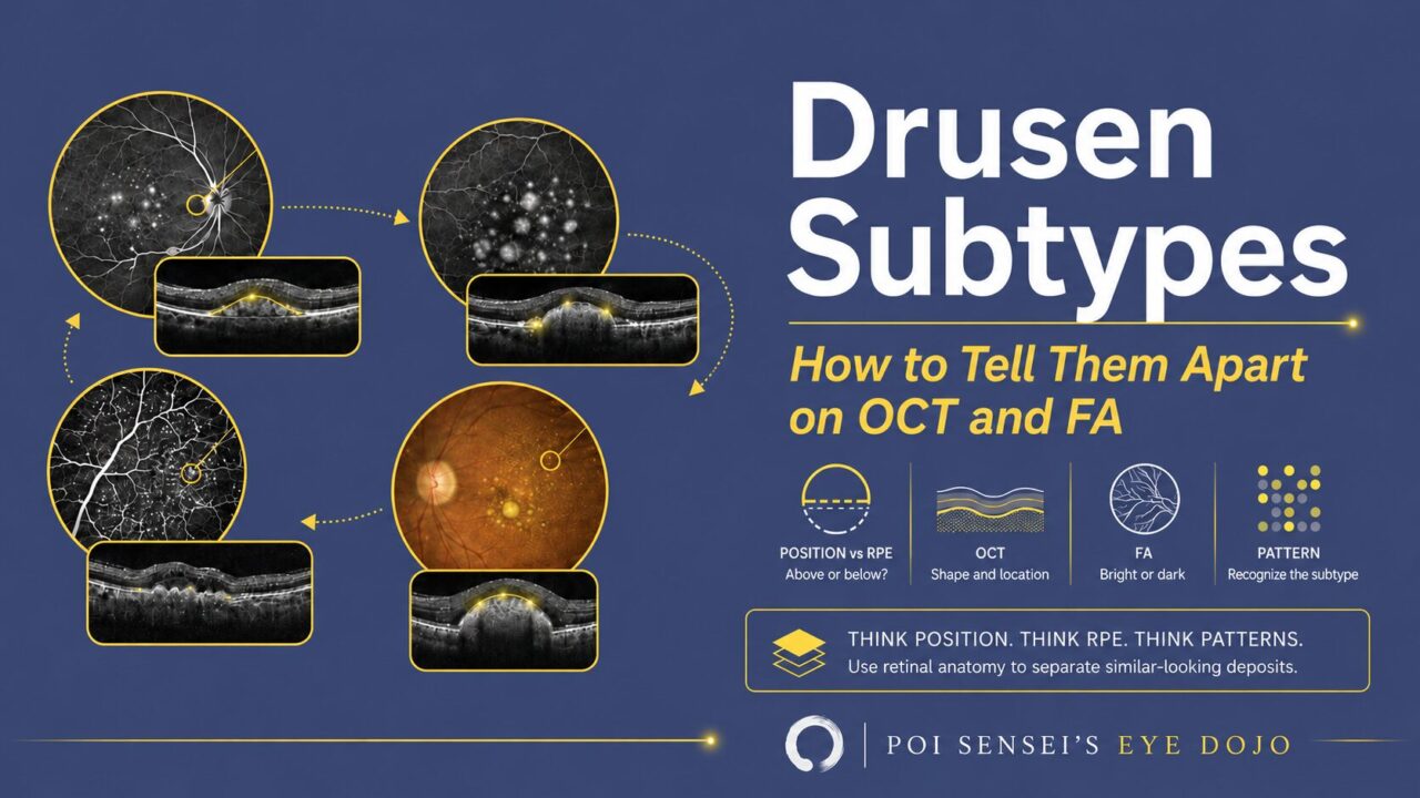
Drusen all look like small yellow-white deposits on a fundus photograph. The subtypes are separated by something you cannot see directly in that photograph: where the deposit sits relative to the retinal pigment epithelium (RPE).
Once you know the position, the imaging follows. You can predict what OCT will show and whether the lesion will be bright or dark on fluorescein angiography (FA), instead of memorizing a table of findings.
Start with size, then describe the deposit
Start with the ordinary drusen you see every day. Two things are being described at the same time — how big the deposit is, and what its border looks like — and the words for them are easy to mix up. Separate them first, then put them back together.
Size comes in three bands, divided at 63 and 125 µm.
- Small — under 63 µm.
- Medium — 63 to 125 µm.
- Large — 125 µm or more.
Shape and border describe the deposit itself. Hard drusen are round and sharply outlined. Soft drusen have indistinct borders that slope into the surrounding retina, and neighboring ones can run together.
The two axes line up, which is why the words are often used interchangeably. Hard drusen are the small ones, under 63 µm. Soft drusen begin at 63 µm, so they fall in the medium and large bands. The 63 µm boundary does double duty: it divides small from medium on the size scale, and it is also the conventional cutoff between hard and soft. That is why you will often see hard and soft defined by size alone — hard is small, soft is 63 µm and up.
Keep the two words apart anyway, because they answer different questions. Size answers how much risk, and the risk rises band by band. Small hard drusen are common in older eyes and, on their own, are not treated as a sign of age-related macular degeneration (AMD). Once the deposits reach soft drusen size, that changes: medium drusen, 63 to 125 µm, already place the eye in the early AMD category, and large drusen, 125 µm or more, are the major risk factor for progression to advanced disease. Soft drusen are not a finding to note and forget — they are the reason to follow the eye.
The border answers a different question: what am I looking at, and it is the part you judge directly at the slit lamp. The two usually agree. When they do not, say both — "medium drusen with sharp borders" describes the lesion better than either word alone.
A practical way to judge size without measuring. A retinal vein at the optic disc margin is about 125 µm across, which is the large-drusen threshold itself. Compare the deposit with that vessel. As wide as the vein or wider means large. Less than half of it means small, which is the hard drusen band.
Then ask where the deposit sits
This is the question that separates the subtypes, and OCT is what answers it. The two landmarks are the RPE and Bruch membrane, described in the retinal anatomy article.
Soft drusen sit behind the RPE, between the RPE basement membrane and the inner collagenous layer of Bruch membrane. On OCT they appear as elevations underneath the RPE.
On FA, soft drusen are hyperfluorescent but do not leak. The bright area stays inside its own borders through the study. If you see true leakage, you are looking at something other than drusen.
Soft drusen also change over time, and the changes are visible on OCT. Adjacent large drusen may become confluent and lift the RPE as a drusenoid pigment epithelial detachment (PED). Drusen can also regress: the material disappears, the elevation collapses, and atrophy of the overlying RPE and photoreceptors may follow. A flattening druse is therefore not automatically good news, and comparing serial scans at the same location is more informative than any single scan.
Subretinal drusenoid deposits (reticular pseudodrusen)
A note on the name first. These were long called reticular pseudodrusen, and many clinicians still use that term. The preferred name is now subretinal drusenoid deposits (SDD). Both names appear in the literature, so it is worth recognizing each.
The name is precise in a useful way. These are drusenoid — they resemble drusen without being drusen. The material differs from soft drusen, and so does the location.
SDD sit on the other side of the RPE. They are deposited on the retinal side, and some cross the external limiting membrane. On a fundus photograph they appear as small punctate yellow-white lesions, often most obvious near the superior arcade.
OCT is where you confirm them. They appear as hyperreflective material in the subretinal space, above the RPE, arranged in a dot-like or ribbon-like pattern, and taller deposits indent the overlying photoreceptor layers. On near-infrared reflectance they appear as hyporeflective lesions against a lighter background, which is where the reticular network is easiest to appreciate.
That position also explains the FA finding in one step. Normally the RPE partly masks choroidal fluorescence, and that background glow is the window effect described in the FA article. SDD sit in front of that glow and block it, so they are hypofluorescent on FA. Their location also helps explain their characteristic appearance on indocyanine green angiography and fundus autofluorescence, though each of those modalities has its own signal source and its own patterns.
SDD have been linked to atrophic AMD, and they are a strong risk factor for retinal angiomatous proliferation (RAP).
Cuticular drusen
The composition resembles ordinary drusen, but the appearance is different: they are numerous, uniform in size, and individually small — roughly 50 to 75 µm.
- Fundus — fine yellow-white spots, often in the posterior pole and nasal to the disc.
- OCT — small deposits under the RPE. Each one is a triangular or dome-shaped elevation with its base on Bruch membrane and its apex toward the retina, and a row of them gives the RPE the sawtooth pattern.
- FA — the many small drusen light up as points of hyperfluorescence, an appearance called stars in the sky.
Cuticular drusen may be accompanied by an acquired vitelliform detachment: yellowish subretinal material at the fovea, seen on OCT as hyperreflective material in the subretinal space. It is worth recognizing, because it is a separate reason for vision to drop in an eye whose drusen alone would not explain it.
Pachydrusen
These accompany a pachychoroid. Across the posterior pole you see scattered yellow-white lesions that are irregular in shape yet have distinct borders, often solitary or in small groups, and they fall into the large drusen size range (125 µm or more). The distribution is wide and tends to spare the central macula, which is the opposite of the way soft drusen cluster there.
On OCT they sit under the RPE like ordinary drusen, and the choroid beneath is thick. That thick choroid is the point: pachydrusen belong with the pachychoroid diseases such as central serous chorioretinopathy and polypoidal choroidal vasculopathy, rather than with the drusen of typical AMD.
Side by side
| Subtype | Position vs RPE | Size | OCT | FA |
|---|---|---|---|---|
| Hard drusen | Under the RPE | Small, under 63 µm | Small discrete sub-RPE elevations | Hyperfluorescent, no leakage |
| Soft drusen | Under the RPE | Medium to large, 63 µm or more | Broad sub-RPE elevations, may become confluent as a drusenoid PED | Hyperfluorescent, no leakage |
| SDD (reticular pseudodrusen) | Above the RPE, subretinal | Small, numerous | Hyperreflective subretinal dots or ribbons | Hypofluorescent by blockage |
| Cuticular drusen | Under the RPE | 50 to 75 µm, numerous | Sawtooth RPE, triangular elevations | Stars in the sky |
| Pachydrusen | Under the RPE | 125 µm or more | Sub-RPE deposits over a thick choroid | Hyperfluorescent, no leakage |
In short
- Position relative to the RPE is the organizing fact. Behind it, in front of it, or under it in fine deposits.
- Hard and soft describe the border, but they track size: hard drusen are under 63 µm, soft drusen are 63 µm or more. The border names the deposit, the size says how much risk it carries.
- Soft drusen sit behind the RPE: hyperfluorescent on FA, without leakage. Follow them over time for confluence, drusenoid PED and collapse.
- SDD, formerly reticular pseudodrusen, sit in front of the RPE: hyperreflective on OCT, hypofluorescent on FA by blockage, and their location shapes how they look on ICGA and autofluorescence too.
- Cuticular drusen give a sawtooth RPE on OCT and stars in the sky on FA, and may carry an acquired vitelliform detachment.
- Pachydrusen are large, irregular and scattered, spare the central macula, and come with a thick choroid.
- Size sorts the risk, and it rises band by band: small hard drusen are not a sign of AMD on their own, medium drusen already fall in the early AMD category, and large drusen are the major risk factor for progression. The retinal vein at the disc margin is a usable 125 µm ruler.



Which retinal layer makes which wave, the three abnormal patterns, and the dark-adaptation trap to check before you report “negative” — on one page, by email. One email when I publish something new, about once a month. Unsubscribe anytime.