Hess Chart Interpretation: Which Muscle Is Weak, and Is It Really a Palsy?
What a Hess chart can tell you
A Hess chart answers two questions, in this order: which eye is not moving fully and in which direction, and — the one that matters more — whether that pattern makes sense as a nerve palsy. Ask them the wrong way round and you will call an esotropia a palsy, or a myasthenic eye a third nerve.
The Hess screen is standard in the UK, Europe and Japan. In North America the same chart usually comes from the Lancaster red-green test, and it reads the same way.
This is the order I use.
- Compare the two fields. The smaller field is the paretic eye.
- Find the short direction in the smaller field. That names the underacting muscle.
- Check the yoke muscle in the other eye. If it overacts, the underaction is a true palsy. If it does not, the short direction is only apparent.
- Ask whether the pattern follows a nerve. If it does not, think muscle or neuromuscular junction, or a central lesion.
The two charts below are real, from Japan’s national examination for certified orthoptists. The original labels are in Japanese, so I have added the English abbreviations. Everything in green is mine; everything else is the original chart.
Case 1: two days of double vision
A 65-year-old man with diabetes has had double vision for two days. The anterior segment, media and fundus are normal.
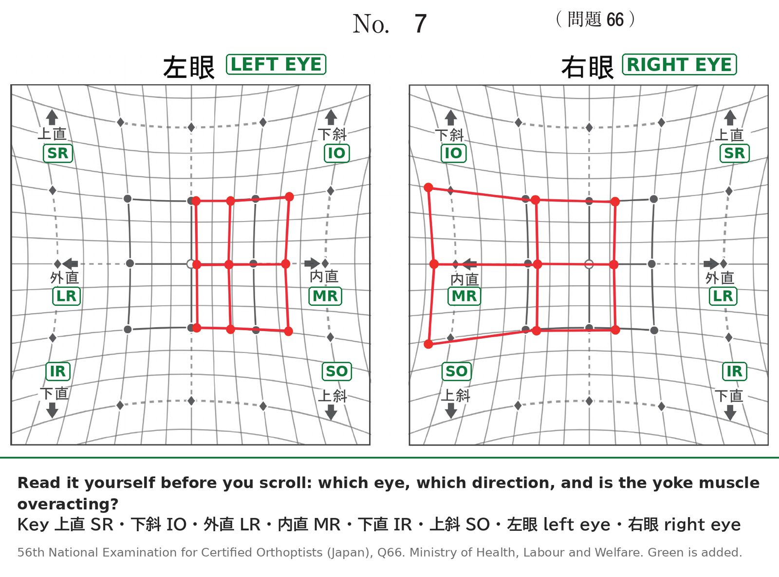
Work through steps 1 to 3 before you read on.
Reading it, step by step
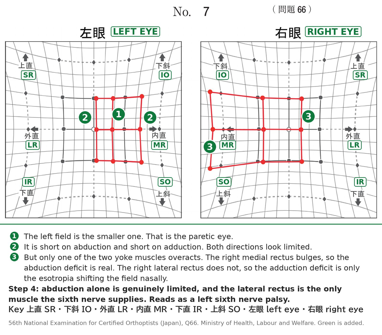
① The left field is the smaller one, so the left eye is the paretic eye.
② Both horizontal directions look short. The left field does not reach the fixation point on abduction, and it falls short on adduction as well. Stop reading here and you would call this a combined horizontal palsy. If you need reminding which muscle does what, the actions are laid out in our guide to extraocular muscle actions.
③ Now the yoke muscles, which is the step beginners skip. A paretic muscle makes its yoke in the fellow eye overact, because both get the same innervation (Hering’s law). On the chart that shows as the fellow field bulging outward — so check both directions. The yoke of the left lateral rectus is the right medial rectus, and the right field bulges a long way nasally. The yoke of the left medial rectus is the right lateral rectus, and on the temporal side it does not bulge at all. Only one of the two is overacting. The abduction deficit is real; the short adduction is the esotropia shifting the whole field nasally, and nothing more.
④ So only abduction is genuinely limited — and that fits one nerve. The lateral rectus is the only muscle the sixth nerve supplies, so an isolated abduction deficit sits exactly inside its territory. The chart reads as a left sixth nerve palsy, not a combined horizontal palsy. In a 65-year-old diabetic with a two-day history, a microvascular cause is the first one to suspect.
Step 4 in full: nerve, muscle or junction?
Case 1 made step 4 look easy, because the deficit landed inside one nerve’s territory. It is often not that clean. There are three places the problem can be: the nerve, the muscle, or the junction between them.
A nerve palsy limits what that nerve supplies, and nothing else. Sixth: abduction. Third: adduction, elevation and depression together, usually with ptosis. Fourth: depression in adduction.
A muscle problem can be a tight muscle or a weak one, and the two get conflated. Thyroid eye disease is the classic tight muscle. It will not lengthen, so it limits gaze away from its own action: an involved inferior rectus limits elevation. A muscle trapped in a fracture, and a congenital tether such as Brown syndrome, behave the same way.
Orbital myositis does not. The inflamed muscle is weak before it is tight — it goes paretic in the first couple of weeks and only later stiffens into a restriction, and when the limitation is measured it is more often in the direction the muscle itself pulls than in the opposite one[1][2]. So an inflamed lateral rectus can give you an abduction deficit that looks exactly like a sixth nerve palsy. IgG4-related orbital disease sits in the same group.
Several muscles limited across different nerve territories still argue against a nerve. One muscle is a genuine mimic, and the chart will not settle it. Forced duction separates tight from weak, and pain on eye movement points at myositis.
A junction problem is myasthenia. The muscles are weak rather than tight, the pattern can copy any nerve, and it is fatigable: worse in the evening, different at every visit. If the chart changes between two visits without the story changing, think myasthenia.
So after the three steps I ask: do the limited movements match one nerve’s territory? An isolated adduction deficit does not fit the third nerve, which should take elevation and depression with it — think internuclear ophthalmoplegia. One or two of the three third-nerve movements: think myasthenia, thyroid or myositis first. All three: third nerve palsy.
Case 2: diplopia after a fall
A 29-year-old man hit a street tree while cycling and fell. His nose bled, and he then saw double. Corrected acuity is 1.2 in each eye.
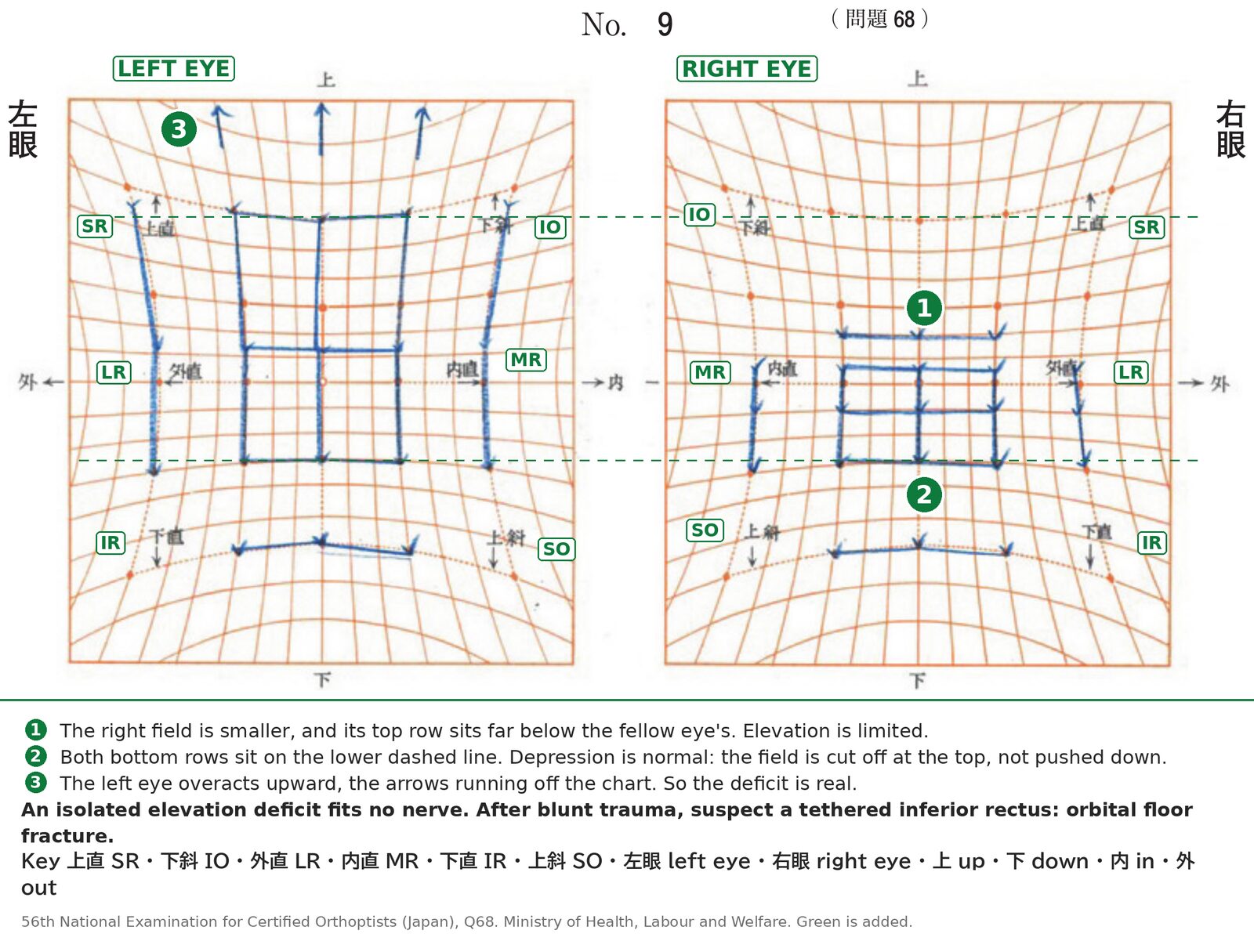
① The right field is the smaller one, so the right eye is the one to look at.
② Its top row sits far below the fellow eye’s. Elevation is limited.
③ The left eye’s upward points run off the chart as arrows — a large overaction — so the limitation is real.
Then step 4. Elevation is limited and depression is not — the two bottom rows sit on the same line. No single cranial nerve does that. A third nerve palsy would take adduction and depression with it; a fourth limits depression, not elevation.
An isolated elevation deficit after blunt trauma means a tethered globe rather than a denervated one, and the muscle to suspect is an inferior rectus caught in an orbital floor fracture. Forced duction confirms the restriction and CT shows the fracture; the chart only tells you to go and do both.
When it is the third nerve: the pupil
The parasympathetic fibers to the pupil run on the surface of the third nerve. Compression from outside, classically a posterior communicating artery aneurysm, hits them first, so the palsy comes with a dilated pupil and ptosis. Ischemia, as in diabetes, damages the centre and spares the surface, so the pupil is often normal.
An abduction deficit is not a sixth nerve palsy
Case 1 finished by matching one deficit to one nerve, and that is where most of the errors happen. "The eye will not abduct" is a finding. "Sixth nerve palsy" is a conclusion, and the differential in between is wider than the nerve. The mnemonic I use is pseudo-BCG:
- pseudo: orbital inflammatory pseudotumor
- B: blow-out fracture
- C: congenital (Duane retraction syndrome)
- G: Graves disease (thyroid eye disease) and myasthenia gravis
Blow-out fracture is on the list because older patients often do not remember the injury. A medial wall fracture can trap the medial rectus and quietly limit abduction, with none of the drama of case 2.
Myasthenia is the one that catches people out. When it weakens several muscles at once the pattern fits no nerve and you think of it straight away. But it can happen to weaken the lateral rectus and little else, and then it hands you a clean isolated abduction deficit — the same chart as case 1. The chart will not separate them. Ask about fatigue and ptosis, and compare the chart with the next one.
Summary
- Small field = paretic eye. Short direction = underacting muscle. Yoke overaction = true palsy. Without the overaction, the short direction is probably the deviation shifting the field.
- Then ask whether the pattern follows a nerve. Thyroid disease, a trapped muscle, myositis and myasthenia do not respect one nerve — but a single inflamed muscle copies one exactly, so pain and forced duction matter.
- An isolated adduction deficit is not a third nerve palsy. Think internuclear ophthalmoplegia.
- An isolated elevation deficit after trauma is a tethered inferior rectus.
- A third nerve palsy with a normal pupil still needs a work-up[3][4].
- Not being able to abduct is not a sixth nerve palsy. Run pseudo-BCG first, and remember myasthenia can weaken the lateral rectus alone.
References
[1] Siatkowski RM, Capó H, Byrne SF, Gendron EK, Flynn JT, Muñoz M, Feuer WJ. Clinical and echographic findings in idiopathic orbital myositis. American Journal of Ophthalmology. 1994;118(3):343–350. PMID 8085592.
[2] Kang MS, Yang HK, Kim N, Hwang JM. Clinical features of ocular motility in idiopathic orbital myositis. Journal of Clinical Medicine. 2020;9(4):1165. PMID 32325733.
[3] Kissel JT, Burde RM, Klingele TG, Zeiger HE. Pupil-sparing oculomotor palsies with internal carotid-posterior communicating artery aneurysms. Annals of Neurology. 1983;13(2):149–154. PMID 6830174.
[4] Trobe JD. Isolated pupil-sparing third nerve palsy. Ophthalmology. 1985;92(1):58–61. PMID 3974995.
Figure credits
Charts reproduced from the 56th National Examination for Certified Orthoptists (Japan), questions 66 and 68, published by the Ministry of Health, Labour and Welfare. Anything in green is mine.