Internuclear Ophthalmoplegia and One-and-a-Half Syndrome: Localizing a Horizontal Gaze Palsy
One eye that will not adduct. Two eyes that will not look to one side. One eye that can only abduct. Each of these patterns tells you where to look in the brainstem, and you can work it out at the bedside. This article shows the pathway for horizontal gaze once, then shows where the lesion is for each pattern.
Eye movement disorders fall into two groups. Supranuclear disorders sit on the path from the brain down to the nuclei of cranial nerves III, IV and VI. Peripheral disorders sit between the nucleus and the muscle. Here we stay above the nerves, with horizontal gaze only.
- Ask the patient to look right and left. Which eye fails, and in which direction?
- One eye fails to adduct: think of the MLF (medial longitudinal fasciculus) on that side (internuclear ophthalmoplegia).
- Both eyes fail to look to one side: think of the PPRF (the horizontal gaze center in the pons) on that side (horizontal gaze palsy).
- Only one eye can still move horizontally, and only outward: one-and-a-half syndrome.
- Check convergence every time: bring a target toward the nose and watch whether the eye that would not adduct now adducts.
The pathway for looking to the right
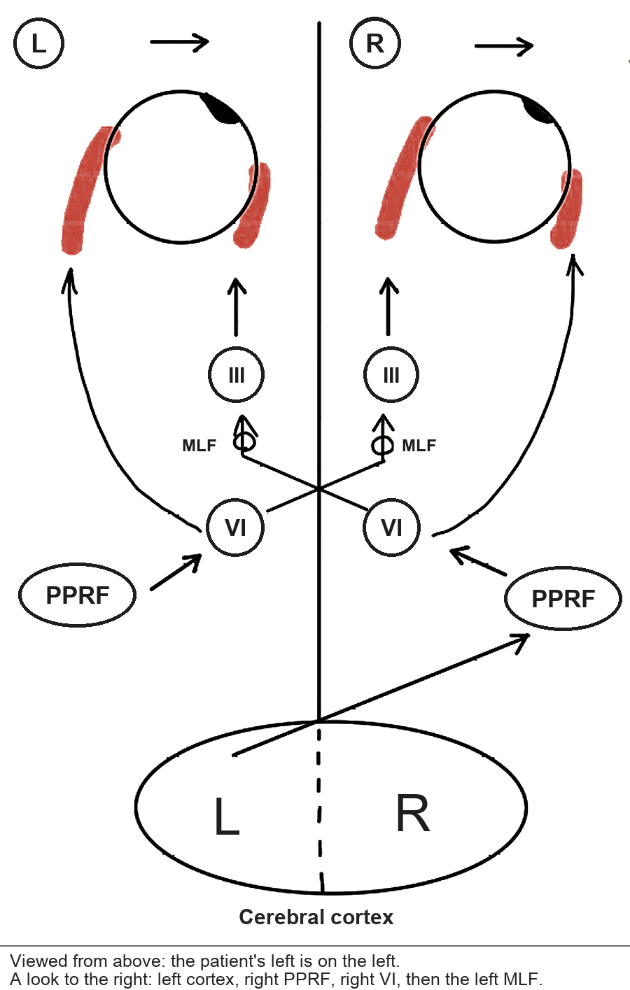
Follow the signal for a look to the right.
- It starts in the left cerebral cortex (the frontal eye field).
- It crosses to the right PPRF (paramedian pontine reticular formation), the horizontal gaze center in the pons.
- The right PPRF drives the right abducens nucleus, and the right lateral rectus contracts.
- The right abducens nucleus also sends a second set of neurons across the midline. They run up in the left MLF (medial longitudinal fasciculus) to the left oculomotor nucleus, and the left medial rectus contracts[1].
This is the base for everything below, so it is worth learning well. The side of the MLF is the part people mix up. One way to hold it:
- The MLF is on the same side as the medial rectus that contracts.
- The PPRF is on the same side as the lateral rectus that contracts, which is also the direction of gaze.
Now let us see what happens when each part is damaged.
Reading each lesion
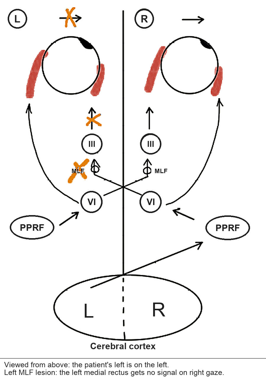
Left MLF: internuclear ophthalmoplegia (INO)
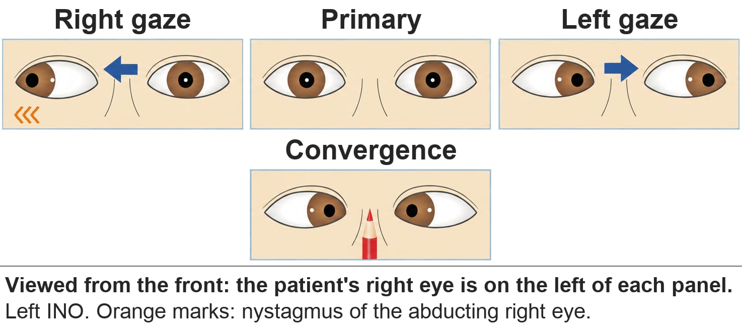
On right gaze, the left eye does not adduct. On left gaze, everything is normal. So when a patient presents with acute double vision and an adduction deficit in one eye only, think of the MLF, for example a small infarct in the pons.
Two more signs complete the picture. The abducting right eye shows nystagmus on right gaze. And when the patient looks straight ahead, the eyes are aligned. The INO is named for the side of the adduction deficit, which is the side of the lesion[1].
The most useful test is convergence. In an isolated MLF lesion, the eye that would not adduct on side gaze adducts on convergence. The medial rectus and the third nerve are healthy, and convergence reaches the oculomotor nucleus by a different route. That test helps separate an INO from a medial rectus or third nerve problem. A large lesion can take convergence too[1].
A mild INO can be subtle. Ask the patient to look quickly from side to side: the adducting eye arrives late[2]. The common causes are demyelination (multiple sclerosis) and stroke[1].
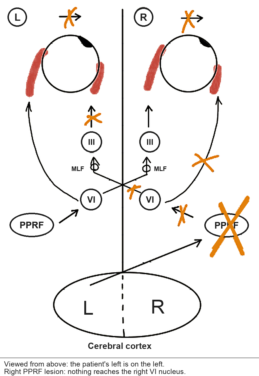
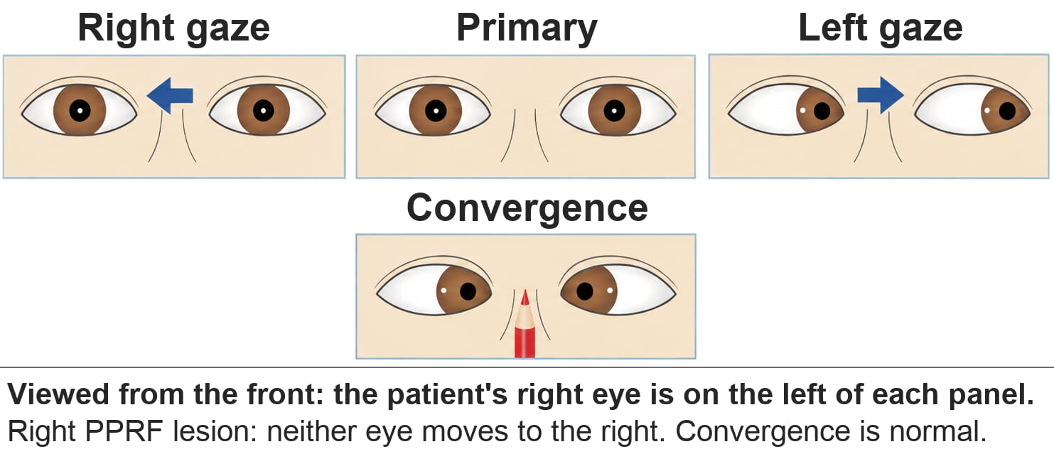
Right PPRF: horizontal gaze palsy
On right gaze, neither eye moves: the right eye does not abduct and the left eye does not adduct. Left gaze is normal. Convergence is preserved here too.
A lesion of the right abducens nucleus gives the same picture when the patient tries to look right, not a sixth nerve palsy. The nucleus drives both the right lateral rectus and the crossing fibers to the left medial rectus, so losing it takes out the whole gaze to that side[2].
One-and-a-half syndrome
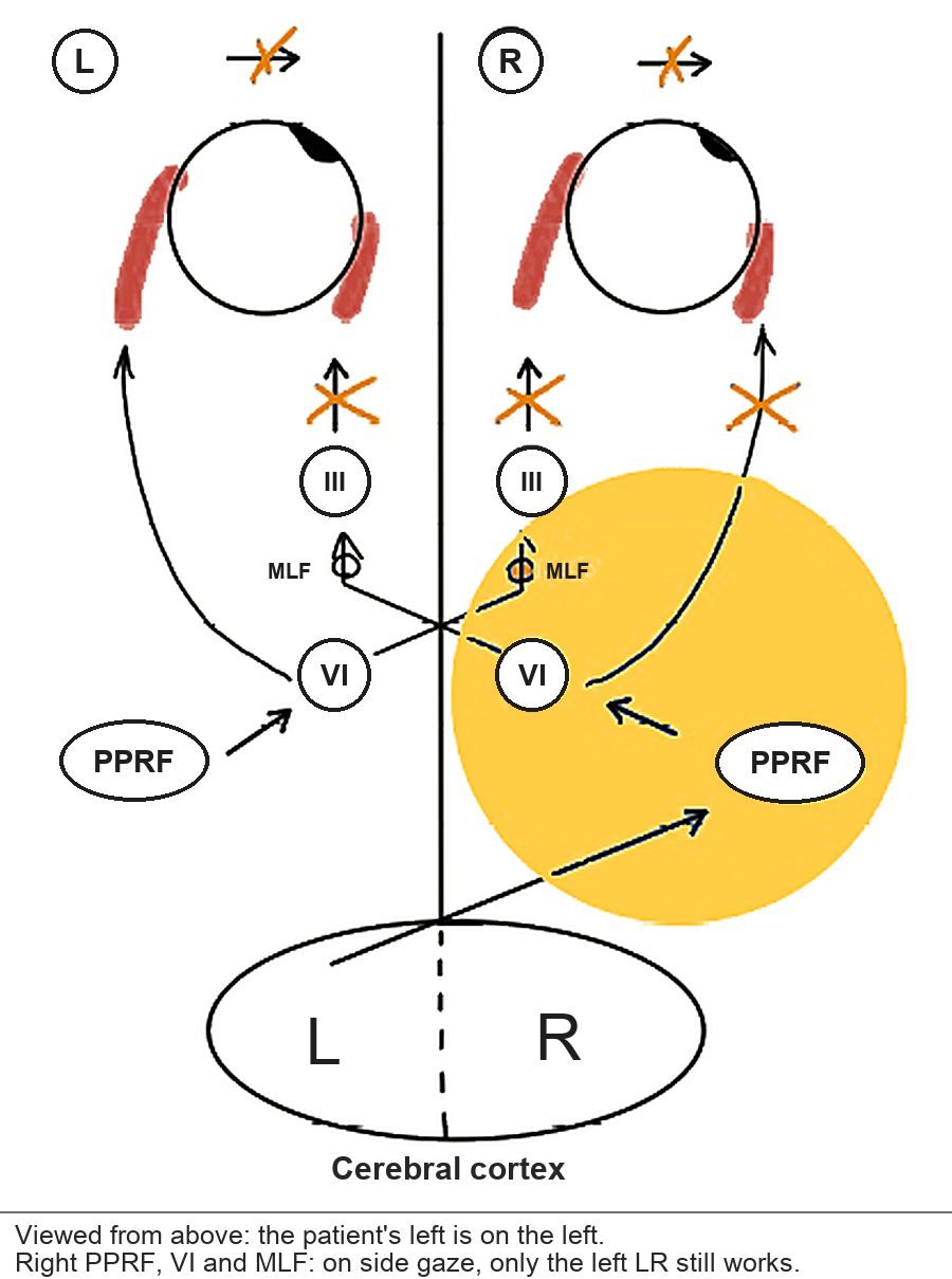
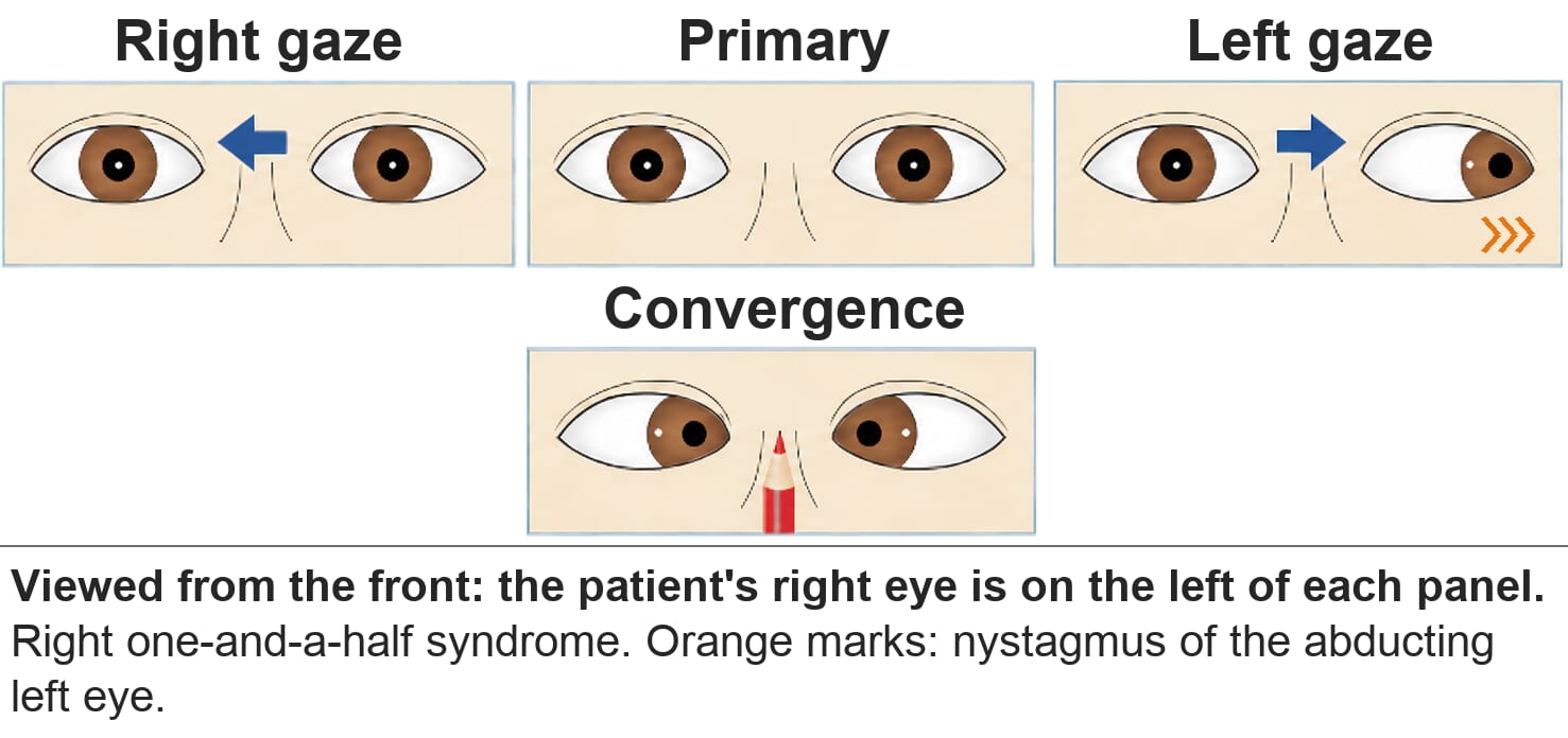
Now suppose a larger lesion takes out the right PPRF and the right MLF together.
- On right gaze, neither eye moves (the gaze palsy).
- On left gaze, the left eye abducts, but the right eye does not adduct (the right INO).
Of the two eyes’ four horizontal movements, only one is left: abduction of the left eye, often with nystagmus[1]. Gaze to the right is lost in both eyes (the "one"), and gaze to the left is lost in the right eye (the "half"), hence the name one-and-a-half syndrome. Convergence is usually still possible. The common causes are multiple sclerosis and brainstem stroke, followed by tumors[2].
Left cerebral cortex
This one is a bonus. A large lesion of the left hemisphere, such as a left middle cerebral artery infarct, also stops the patient looking to the right, like a right PPRF lesion. Because the pull to the right is gone, the eyes rest toward the left, toward the side of the lesion. This is the conjugate deviation that comes with a large stroke.
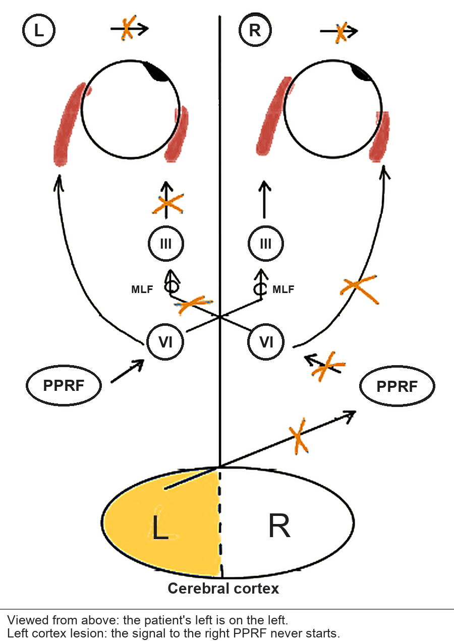
A seizure does the opposite. A left hemisphere seizure overstimulates the pathway, so the eyes turn to the right, away from the lesion[1].
The same logic works in the pons: when the right PPRF is lost, the eyes tend to rest toward the left, away from the lesion.
Summary
- Learn the pathway for one direction: cortex, opposite PPRF, abducens nucleus, then crossing fibers in the MLF to the opposite medial rectus.
- One eye that will not adduct, with nystagmus of the abducting eye, is an INO on the side of the eye that will not adduct.
- Both eyes that will not look to one side point to the PPRF or abducens nucleus on that side.
- One-and-a-half syndrome leaves only abduction of the eye on the other side from the lesion.
- Convergence is usually preserved in all of them. Rule out myasthenia before you call it an INO.
Figure credits
The pathway diagrams are my own drawings. The eye-position drawings were made with an image generator, using as a style model the figure from the 53rd National Examination for Certified Orthoptists (Japan), afternoon session, question 66 (booklet No. 4), published by the Ministry of Health, Labour and Welfare. The labels on the eye-position drawings are mine.
References
[1] Salmon JF. Kanski’s Clinical Ophthalmology: A Systematic Approach. 10th ed. Elsevier; 2025. Chapter 19, Neuro-ophthalmology: supranuclear disorders of ocular motility; abnormalities of horizontal gaze.
[2] Yanoff M, Duker JS, eds. Ophthalmology. 5th ed. Elsevier; 2019. Chapters 9.14, Disorders of supranuclear control of ocular motility, and 9.15, Nuclear and fascicular disorders of eye movement.