Peripheral Corneal Thinning: Pellucid, Terrien and Mooren
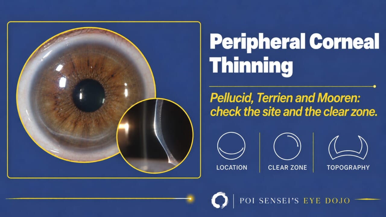
Several diseases thin the peripheral cornea. This article shows how to tell them apart: pellucid marginal degeneration from keratoconus on topography, and from Terrien degeneration, Mooren ulcer, marginal keratitis and keratoglobus at the slit lamp.
- Is the eye red and painful, and does the thinning stain with fluorescein? If so, think of an ulcer, not a degeneration. Rule out infection first.
- If it is an ulcer, is there clear cornea between the lesion and the limbus? A clear zone suggests marginal keratitis. No clear zone suggests an autoimmune ulcer.
- If the eye is quiet, where is the thinning? An inferior band suggests pellucid marginal degeneration. Superior thinning with lipid and vessels suggests Terrien degeneration. Thinning of the whole cornea suggests keratoglobus.
- On topography, look for a crab-claw pattern. Then check the thickness map.
Pellucid marginal degeneration
Pellucid marginal degeneration (PMD) is a slowly progressive thinning and protrusion of the inferior cornea. It causes irregular, against-the-rule astigmatism. It usually appears between the ages of 20 and 40, and both eyes are affected.
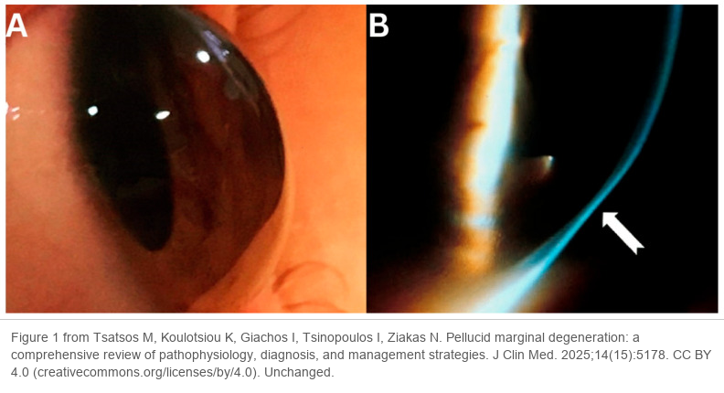
The thinning is a crescent-shaped band from about 4 to 8 o’clock, about 1 mm inside the limbus. The cornea above it is clear and bulges forward. Seen from the side, the cornea protrudes just above the thinnest band[1].
PMD looks like keratoconus, so these two need to be told apart first. Other diseases also thin the peripheral cornea: peripheral ulcers, Mooren ulcer and Terrien marginal degeneration.
Pellucid marginal degeneration vs keratoconus
The main differences
Keratoconus thins and protrudes in the center or just below it, and the thinnest point sits at the cone. PMD does so in the inferior periphery. Keratoconus also starts earlier, around puberty, while PMD starts later.
| Keratoconus | PMD | |
|---|---|---|
| Thinning | Center or just below | Inferior band |
| Onset | Puberty | Age 20 to 40 |
| Fleischer ring, Vogt striae | Common | Usually absent |
They also have things in common. Both are treated with rigid contact lenses. Both can develop acute hydrops, which leaves a scar, although hydrops is less common in PMD.
Topography: the crab-claw pattern
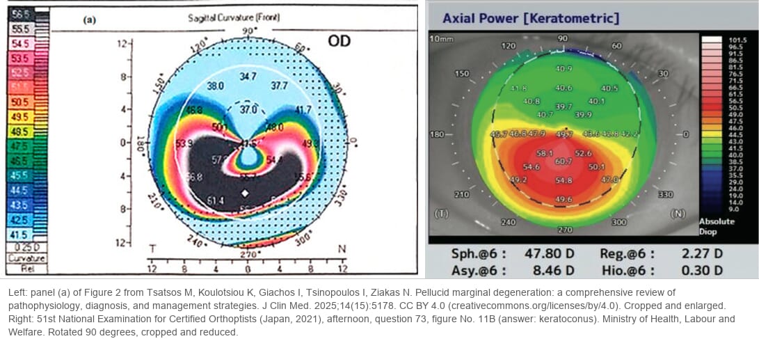
Corneal topography is the most useful test to tell the two apart. This is a curvature map: blue is flat and red is steep. In PMD, a flat zone runs vertically through the center, and a steep zone wraps around it from below. The shape looks like a crab’s claw, so it is called the crab-claw appearance (also "kissing doves"). It is characteristic of PMD.
In keratoconus, the steep area gathers into a round cone in or just below the center.
Pellucid marginal degeneration vs peripheral ulcers
Because PMD thins the inferior cornea, it also has to be told from Mooren ulcer and Terrien marginal degeneration. The biggest difference from Mooren ulcer is that PMD is not inflammatory. The eye is white, the epithelium is intact and there is no pain. Terrien degeneration is also usually quiet, so look for its lipid and vessels.
Mooren ulcer
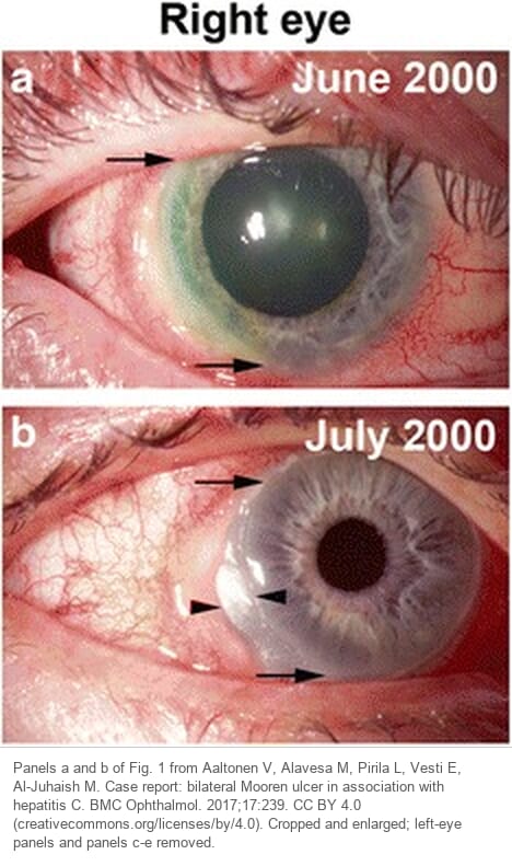
Mooren ulcer is an autoimmune ulcer of the peripheral cornea in a patient with no infection or systemic disease that explains the ulcer. Pain is severe, the eye is red, and the ulcer has an epithelial defect that stains with fluorescein. The ulcer starts at the limbus and spreads around it and toward the center. The edge on the central side is undercut, with a white infiltrate. None of these signs appear in PMD.
Mooren ulcer does not spread into the sclera. If scleritis is present, think of an ulcer caused by systemic disease instead[3].
Terrien marginal degeneration
Terrien marginal degeneration is a peripheral thinning that can start at any age, from young adults to the elderly. Both eyes are usually affected, but often unequally. The cause is unknown.
It usually begins superiorly with fine opacities in the peripheral stroma. Over years, stromal opacity, superficial vessels and thinning slowly spread parallel to the limbus. Lipid deposits collect on the central side of the thin area, so the central edge shows a yellow-white line. About 20% of patients develop a pseudopterygium. Like PMD, it causes against-the-rule astigmatism.
At a glance it can look like Mooren ulcer. Three points tell them apart:
- The epithelium is intact. Fluorescein shows no defect.
- It progresses slowly, over years. Mooren ulcer progresses faster.
- The eye is usually quiet, with no conjunctival injection and no infiltrate. A minority of patients have episodes of episcleritis or scleritis, so an episode does not rule it out[3].
There is no treatment that stops it. Glasses or contact lenses correct the astigmatism, steroids are sometimes used, and severe thinning may need a lamellar graft.
The clear zone: marginal keratitis vs autoimmune ulcers
Ulcers of the peripheral cornea fall into two groups.
Bacterial hypersensitivity. The bacteria do not invade the cornea. The cornea reacts to an infection of the eyelid. Staphylococcus aureus blepharitis causes marginal (catarrhal) keratitis. These lesions are mainly infiltrates, and the ulcer stays shallow.
Autoimmune. This group includes peripheral ulcerative keratitis (PUK) with rheumatoid arthritis or granulomatosis with polyangiitis (formerly Wegener granulomatosis), and Mooren ulcer. The infiltrate is mild, but the ulcer is deep. It keeps deepening and can perforate.
The key sign to tell the two groups apart is the clear zone: a strip of normal cornea between the lesion and the limbus. It shows where the attack comes from.
- Autoimmune ulcers have no clear zone. The cornea has no blood vessels, so immune cells arrive from the vessels at the limbus. They cannot skip the limbus and start the attack further in, so the ulcer begins at the limbus and stays joined to it.
- Marginal keratitis has a clear zone. The trigger is toxins from the bacteria on the lid margin, not the limbal vessels. The lesion forms where the lid margin rests on the cornea, so it is not connected to the limbus.
Column: LDAK, an ulcer that comes from the tear duct
In 2022 a group in Japan described lacrimal drainage pathway disease-associated keratopathy (LDAK): a noninfectious corneal ulcer caused by chronic dacryocystitis or canaliculitis[4]. In their 13 patients the ulcer was usually peripheral, mostly inferior or nasal, with few infiltrates, so it can look like Mooren ulcer. Seven of the 13 perforated.
The clue is in the tear duct, not the cornea: heavy discharge and a red, swollen punctum. The ulcers did not improve with antibiotic and steroid drops, but once the tear duct was treated they healed in about two weeks and did not recur. It is not only a Japanese disease. A similar case, PUK from chronic canaliculitis, was reported from Taiwan[5].
The mechanism is unclear. Toxins from the bacteria, or enzymes released by the inflamed tear duct, are suspected.
Keratoglobus: thinning of the whole cornea
Keratoglobus is a rare bilateral disease in which the whole cornea thins and bulges forward like a globe. It is often present soon after birth. Rarely it is acquired in adulthood.
It looks quite different from keratoconus and PMD. The corneal diameter is normal, but the stroma is thinned to one-third to one-fifth of normal, most of all in the periphery. Because the whole cornea protrudes, its curvature becomes steep, which causes high myopia and astigmatism.
It progresses little, but the cornea is weak. Descemet membrane can tear (acute hydrops), and the cornea can rupture after minor trauma. Protect these eyes from injury.
Summary
- PMD is an inferior band of thinning in a quiet eye, with against-the-rule astigmatism and onset in adulthood.
- On topography PMD shows a crab-claw map. Inferior keratoconus can look the same, so check the thickness map.
- A red, painful eye with an epithelial defect means an ulcer. A clear zone points to marginal keratitis. No clear zone points to an autoimmune ulcer, such as Mooren ulcer or PUK.
- Terrien degeneration starts superiorly, with lipid at the central edge, superficial vessels and an intact epithelium.
- Keratoglobus thins the whole cornea, usually from birth, and the eye must be protected from trauma.
References
[1] Salmon JF. Kanski’s Clinical Ophthalmology: A Systematic Approach. 10th ed. Elsevier; 2025. Chapter 7, Cornea: Peripheral corneal ulceration/thinning; Pellucid marginal degeneration; Keratoglobus.
[2] Vieira IV, Fan VH, Yu CQ. Update on pellucid marginal degeneration. Graefes Arch Clin Exp Ophthalmol. 2026;264(2):325–335. PMID 41212223.
[3] Yanoff M, Duker JS. Ophthalmology. 5th ed. Elsevier; 2019. Chapter 4.17, Noninfectious keratitis: Mooren’s ulcer; Terrien’s marginal degeneration.
[4] Inoue H, Toriyama K, Ikegawa W, et al. Clinical characteristics of lacrimal drainage pathway disease-associated keratopathy. BMC Ophthalmol. 2022;22:353. PMID 36045355.
[5] Hsieh HH, Shen EP. Peripheral ulcerative keratitis secondary to chronic Citrobacter koseri canaliculitis. Taiwan J Ophthalmol. 2023;13(3):376. PMID 38089517.
Figure credits
Slit-lamp photographs and the left curvature map: Figure 1 and panel (a) of Figure 2 from Tsatsos M, Koulotsiou K, Giachos I, Tsinopoulos I, Ziakas N, J Clin Med 2025;14(15):5178, licensed under CC BY 4.0. Figure 1 unchanged. Figure 2 cropped to panel (a) and enlarged.
Keratoconus map: 51st National Examination for Certified Orthoptists (Japan, 2021), afternoon, question 73, figure No. 11B, published by the Ministry of Health, Labour and Welfare. Rotated 90 degrees, cropped and reduced.
Mooren ulcer photographs: panels a and b of Fig. 1 from Aaltonen V, Alavesa M, Pirilä L, Vesti E, Al-Juhaish M, BMC Ophthalmol 2017;17:239, licensed under CC BY 4.0. Cropped and enlarged; the left-eye panels and panels c to e removed.