Occult Macular Dystrophy (Miyake Disease): OCT and ERG Findings
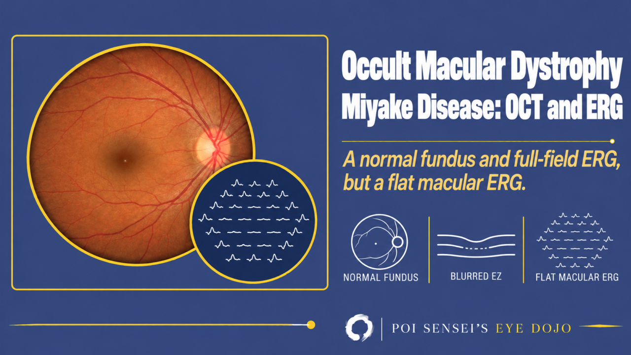
A patient’s vision has been falling slowly in both eyes. The fundus, the angiogram and even the full-field ERG are normal. Think of occult macular dystrophy. This article explains what it is, and how to find it with OCT and the multifocal ERG.
- Vision falls slowly in both eyes.
- The fundus and the fluorescein angiogram look normal.
- The full-field ERG is normal.
- The multifocal ERG is reduced only at the macula.
What is occult macular dystrophy?
Occult macular dystrophy is a macular dystrophy in which macular function falls, but the fundus photograph, fluorescein angiography and fundus autofluorescence show no clear abnormality. "Occult" means hidden from sight[1].
Symptoms
Vision falls slowly in both eyes, while the peripheral visual field stays normal. Corrected visual acuity rarely falls below 20/200 (6/60).
Onset varies widely, from about age 10 to over 60[2].
Classification
Occult macular dystrophy has several types, and the names are confusing.
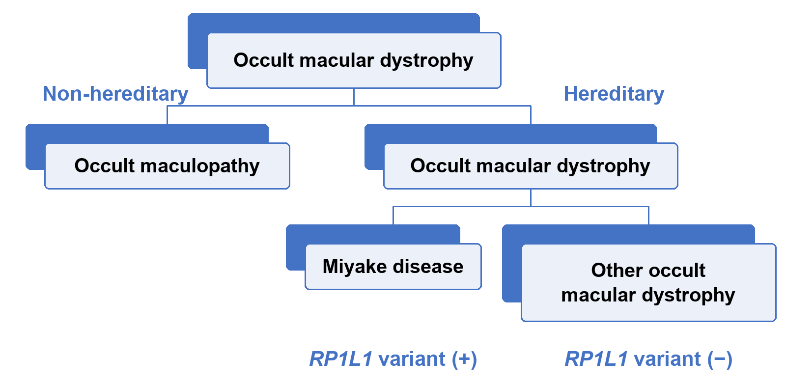
Cases are either hereditary or non-hereditary. Strictly, only hereditary cases are called occult macular dystrophy, and non-hereditary cases are called occult maculopathy. In practice, the name is often used for both. Among the hereditary cases, those with a variant in the RP1L1 gene are called Miyake disease. Miyake disease is autosomal dominant.
Fundus
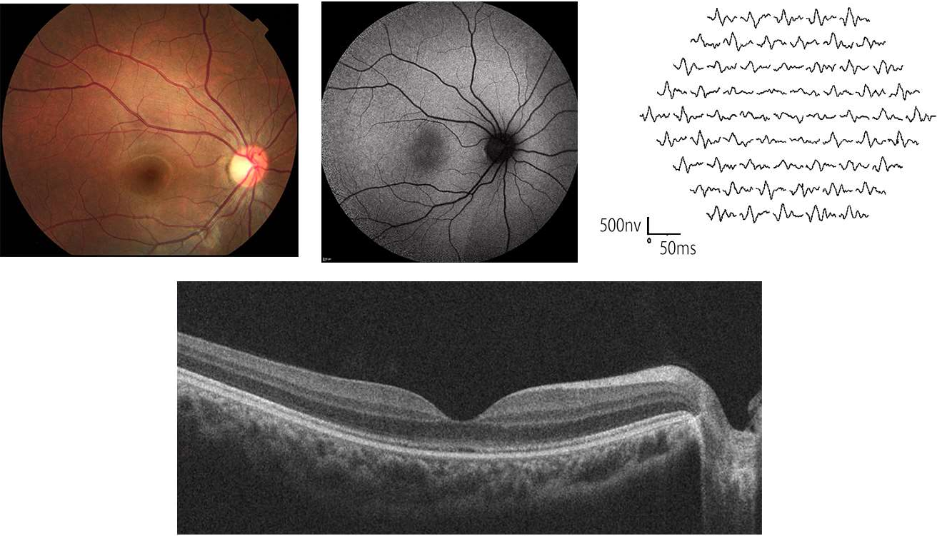
The fundus photograph and fluorescein angiography are normal. Fundus autofluorescence is usually normal too.
OCT
The ellipsoid zone (EZ) and the interdigitation zone (IZ) are two bright lines on OCT that come from the photoreceptors. In occult macular dystrophy, OCT can show a blurred EZ and a missing IZ at the fovea. The retinal pigment epithelium stays normal. These OCT changes are typical of Miyake disease and uncommon in occult maculopathy[2].
ERG findings
Only the macula is affected, and the peripheral retina is normal. The full-field ERG records the whole retina, and the macula is only a small part of it, so the full-field ERG stays normal. The multifocal ERG and the focal macular ERG are reduced only at the macula. This finding confirms the diagnosis[1].
| ERG | Occult macular dystrophy |
|---|---|
| Full-field ERG | Normal |
| Multifocal ERG | Reduced at the center |
| Focal macular ERG | Reduced |
You do not need to think hard about reading a multifocal or focal macular ERG. Where the waveforms are small, that part of the retina is abnormal.
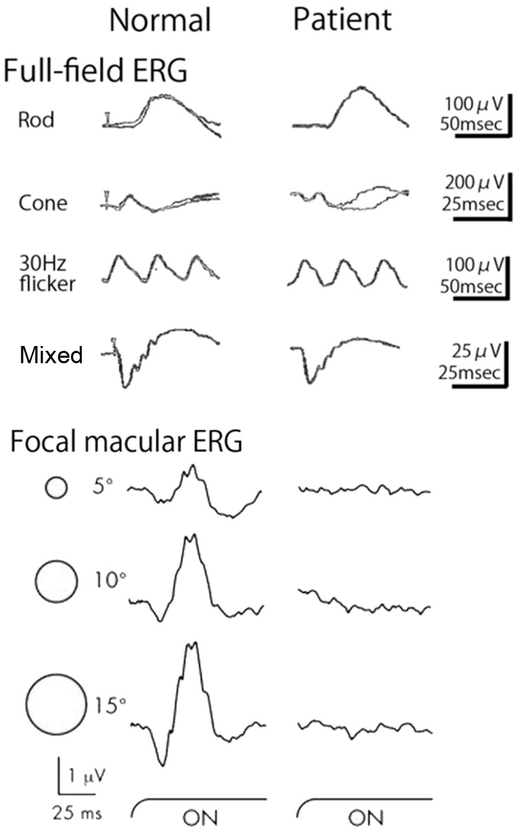
Yozo Miyake developed the focal macular ERG at Nagoya University in Japan and used it to report this disease in 1989. That is why the disease carries his name[1]. Japanese ophthalmologists know it well, and it has come up many times on the Japanese board exam. A pattern ERG can also show the macular dysfunction[2].
Treatment
There is no treatment. Magnifiers and other low vision aids help with reading.
Summary
- Occult macular dystrophy slowly lowers vision in both eyes, with a normal fundus and angiogram.
- Non-hereditary cases are occult maculopathy. Hereditary cases with an RP1L1 variant are Miyake disease.
- OCT: a blurred EZ and a missing IZ at the fovea, typical of Miyake disease.
- ERG: the full-field ERG is normal, but the multifocal and focal macular ERGs are reduced at the macula.
Figure credits
Fundus, autofluorescence, OCT and multifocal ERG: Figure 1 from Fujinami-Yokokawa Y, Yang L, Joo K, et al. Genes 2023;14(10):1869 (https://doi.org/10.3390/genes14101869), licensed under CC BY 4.0. Four panels of the top row cropped and rearranged, without the visual field and the row label.
Full-field and focal macular ERG: Figure 16 from Miyake Y. Int J Mol Sci 2025;26(11):5166 (https://doi.org/10.3390/ijms26115166), licensed under CC BY 4.0. The row label Bright flash was changed to Mixed.
References
[1] Miyake Y. Review of four refined clinical entities in hereditary retinal disorders from Japan. Int J Mol Sci. 2025;26(11):5166. PMID 40507975.
[2] Fujinami-Yokokawa Y, Yang L, Joo K, et al. Occult macular dysfunction syndrome: identification of multiple pathologies in a clinical spectrum of macular dysfunction with normal fundus in East Asian patients: EAOMD report no. 5. Genes. 2023;14(10):1869. PMID 37895218.
[3] Yanoff M, Duker JS. Ophthalmology. 5th ed. Elsevier; 2019. Chapter 6.9, Retinal electrophysiology.
Which retinal layer makes which wave, the three abnormal patterns, and the dark-adaptation trap to check before you report “negative” — on one page, by email. One email when I publish something new, about once a month. Unsubscribe anytime.