Angioid Streaks: Fundus Signs, the Link to PXE and How to Manage MNV
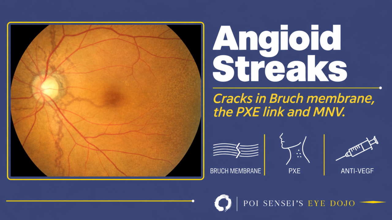
Dark, jagged lines that radiate from the optic disc and lie deeper than the retinal vessels are angioid streaks. They are cracks in Bruch membrane, and new vessels can grow up through the cracks. This article explains why the cracks form, how they look on the fundus and on imaging, which systemic diseases to look for, and how to manage the complications.
- Gray or dark red lines ring the optic disc and branch outward from it, deeper than the retinal vessels.
- The fundus temporal to the macula looks mottled, like orange peel (peau d’orange).
- Macular neovascularization (MNV) appears in a patient younger than the usual age for age-related macular degeneration (AMD).
- The patient has small yellowish bumps on the neck.
What are angioid streaks?
In short, angioid streaks are cracks in Bruch membrane. New vessels from the choroid grow up through the cracks, the way a plant pushes up through a crack in the road. The name means "vessel-like," because the branching lines can look like blood vessels.
Bruch membrane has five layers
Bruch membrane lies between the retinal pigment epithelium (RPE) and the choriocapillaris. From the retinal side, it has five layers:
- Basement membrane of the RPE
- Inner collagenous layer
- Elastic layer
- Outer collagenous layer
- Basement membrane of the choriocapillaris
In angioid streaks, the elastic layer degenerates and calcifies. Bruch membrane becomes thick and brittle, and it cracks. The RPE over a crack becomes atrophic, and new vessels from the choroid can grow through the gap.
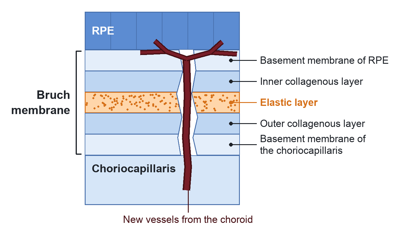
The orange band is the calcified elastic layer. The crack runs through all five layers, and new vessels from the choroid grow through it and spread under the RPE.
Findings
Streaks around the disc
Angioid streaks start at the disc. The lines form a ring around the disc and branch outward from it. They are gray or dark red, with irregular, jagged edges, and they lie deeper than the retinal vessels. They are usually seen in both eyes and slowly grow wider and longer over the years.
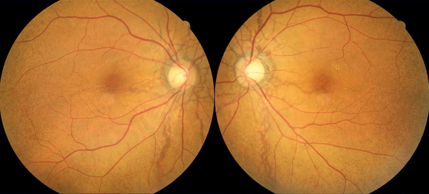
In both eyes, dark lines radiate from the disc, most clearly below it.
Peau d’orange
The other typical sign is peau d’orange ("orange skin"): a mottled, yellowish speckling of the fundus, most obvious temporal to the macula. Where the calcification of Bruch membrane is confluent, the membrane cracks into streaks. Where the calcification is patchy, it gives peau d’orange. Peau d’orange is common in patients with pseudoxanthoma elasticum (PXE), and it often appears before the streaks. In the photographs above, it is the fine, granular look of the fundus temporal to the macula.
Imaging
The streaks are often easier to see on images than with the ophthalmoscope. Red-free and infrared photographs show the lines with more contrast. Fundus autofluorescence (FAF) often shows the streaks over a wider area than you see clinically, and it also shows peau d’orange.
On fluorescein angiography (FA), the streaks look bright, because the background choroidal fluorescence shows through the atrophic RPE (window defects). You do not need FA for the streaks themselves. Use it when you suspect MNV. Indocyanine green angiography (ICGA) shows the streaks more clearly than FA.
Use OCT to check the macula for fluid, blood and new vessels.
Systemic diseases to look for
About half of patients with angioid streaks have a systemic disease. Each of these diseases makes Bruch membrane brittle.
Pseudoxanthoma elasticum (PXE)
By far the most common association is pseudoxanthoma elasticum (PXE), an autosomal recessive disease caused by mutations in the ABCC6 gene. Elastic fibers in the skin, the eye and the arteries calcify and break down. Angioid streaks with PXE are called Grönblad-Strandberg syndrome.
The skin shows small yellowish-white bumps, raised like hives, that are scattered or merge into plaques. They appear on the neck, in the armpits and groin, around the navel, and in the elbow and knee creases.
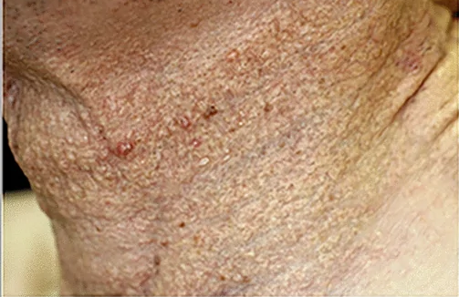
How often PXE lies behind angioid streaks depends on how hard you look. When a group in Kyoto biopsied the skin of patients with angioid streaks, nearly 90% had PXE[1]. In Japan’s national claims data, by contrast, only 4% of patients with angioid streaks had a recorded diagnosis of PXE[2]. In a Tokyo series, patients without a PXE diagnosis had peau d’orange about as often as patients with PXE[3]. So look at the skin yourself, and ask a dermatologist when you are unsure.
PXE also affects the arteries. Once PXE is suspected, the patient needs a check of the heart and blood vessels as well.
Other causes
The other classic associations are Paget disease of bone and hemoglobinopathies such as sickle cell disease and thalassemia. Ehlers-Danlos syndrome still appears on older lists, but a real link is doubtful. In the other half, no associated disease is found.
Complications and treatment
Macular neovascularization
The streaks themselves cause no symptoms. Vision drops when new vessels grow through a crack into the macula. This is macular neovascularization (MNV), often still called choroidal neovascularization. MNV is by far the most common cause of visual loss, and more than 70% of patients eventually develop reduced vision.
In the Tokyo series, about half of the eyes had MNV, and most patients with MNV had it in both eyes[3].
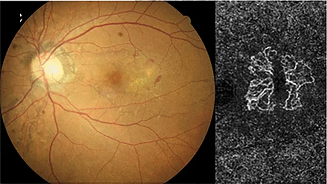
On the left, the MNV sits temporal to the fovea. On the right, OCT angiography shows the network of new vessels.
MNV in angioid streaks starts at a younger age than in AMD. When you see MNV in a younger patient, look around the disc of both eyes for streaks.
Treatment
Treat MNV with intravitreal anti-VEGF injections, as in neovascular AMD. Laser and photodynamic therapy gave poor results. In a series of 39 eyes followed for about six years, vision improved in the first months and then stayed stable, and fewer injections were needed over time[4]. MNV often comes back or appears at a new crack, so follow-up continues for years.
In PXE, some eyes have chronic subretinal fluid that does not come from active MNV. This fluid may not respond to anti-VEGF, so check MNV activity on FA or OCT angiography before you decide to inject.
Streaks without MNV need no treatment. Watch them, and protect the eye.
Same crack, different cause: lacquer cracks in high myopia
High myopia gives a similar picture. One explanation is that as the eye grows longer, Bruch membrane is stretched and splits. These breaks, called lacquer cracks, are shorter and thinner than angioid streaks and cross each other at the posterior pole. Like angioid streaks, they are a risk factor for MNV.
The causes differ, but in both diseases new vessels come through a crack in Bruch membrane. It helps to remember them together.
Summary
- Angioid streaks are cracks in a calcified, brittle Bruch membrane that radiate from the disc.
- Look for peau d’orange, and use red-free, FAF or ICGA when the streaks are faint.
- PXE is the most common cause, so examine the skin, and check the heart and vessels if PXE is suspected.
- Vision is lost through MNV, which is treated with anti-VEGF and often comes back.
- Warn patients to protect the eye from blunt trauma.
References
[1] Nakagawa S, Yamashiro K, Tsujikawa A, Otani A, Tamura H, Ooto S, Yoshimura N. The time course changes of choroidal neovascularization in angioid streaks. Retina. 2013;33(4):825-833. PMID 23114408.
[2] Wada S, Miyake M, Kido A, Kamei T, Hiragi S, Ikeda HO, Hata M, Ueshima H, Tsujikawa A, Tamura H. Epidemiology of angioid streaks and pseudoxanthoma elasticum (2011-2020): a nationwide population-based cohort study. Ophthalmol Sci. 2024;4(2):100370. PMID 37868801.
[3] Maruko R, Maruko I, Hashiya N, Hasegawa T. Clinical characteristics of angioid streaks in Japanese patients. Jpn J Ophthalmol. 2026;70(5):1147-1153. PMID 42397664.
[4] Torres-Costa S, Bernardes J, Mano SS, Medeiros-Pinto J, Abreu AC, Furtado MJ, Silva R, Marques-Neves C, Falcão-Reis F, Carneiro Â, Colaço L, Falcão M. Long-term effect of anti-vascular endothelial growth factor (anti-VEGF) injections in choroidal neovascularization secondary to angioid streaks. J Ophthalmol. 2022;2022:3332421. PMID 35855887.
Which retinal layer makes which wave, the three abnormal patterns, and the dark-adaptation trap to check before you report “negative” — on one page, by email. One email when I publish something new, about once a month. Unsubscribe anytime.