Bietti Crystalline Dystrophy: Crystals, Autofluorescence, ERG and How to Tell It From RP
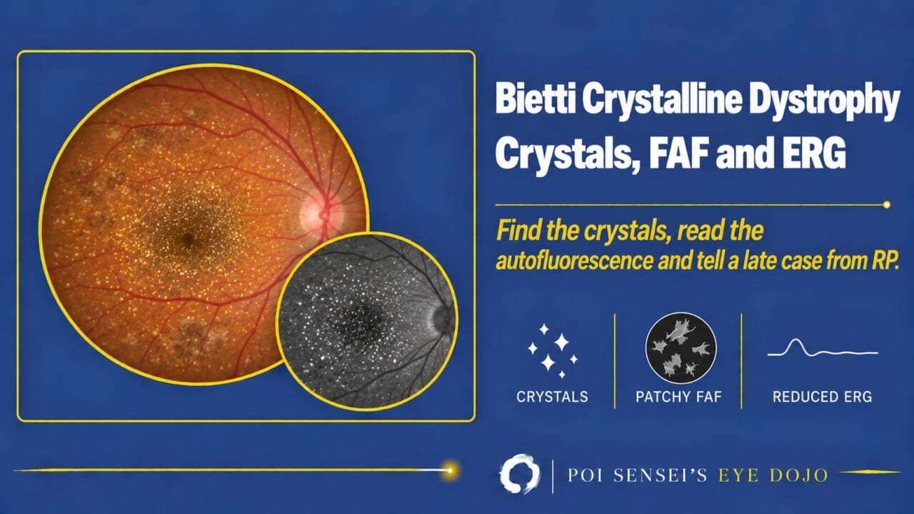
Think of Bietti crystalline dystrophy when a young adult has night blindness and fine glittering crystals in the fundus. This article shows how to read the fundus, autofluorescence and ERG in Bietti crystalline dystrophy, and how to tell a late case from retinitis pigmentosa.
- Look for fine yellow-white crystals over the posterior pole. Near-infrared images show them more clearly.
- Take a fundus autofluorescence image. Are there patchy dark areas?
- Record a full-field ERG. Are rod and cone responses both reduced?
- In a late case the crystals may be few. Then check the retinal vessels and the corneal limbus, and order genetic testing.
What Bietti crystalline dystrophy is
Bietti crystalline dystrophy (BCD) is an inherited disease that causes progressive atrophy of the retinal pigment epithelium (RPE) and choroid. It is autosomal recessive, and the gene is CYP4V2[1].
Who gets it
BCD is rare, but it is more common in Chinese and Japanese people than in other ethnic groups.
Symptoms
Typically, night blindness starts in the 20s or 30s, in men and women alike. In the visual field, sensitivity drops near the center (paracentral scotoma).
Findings
Fundus
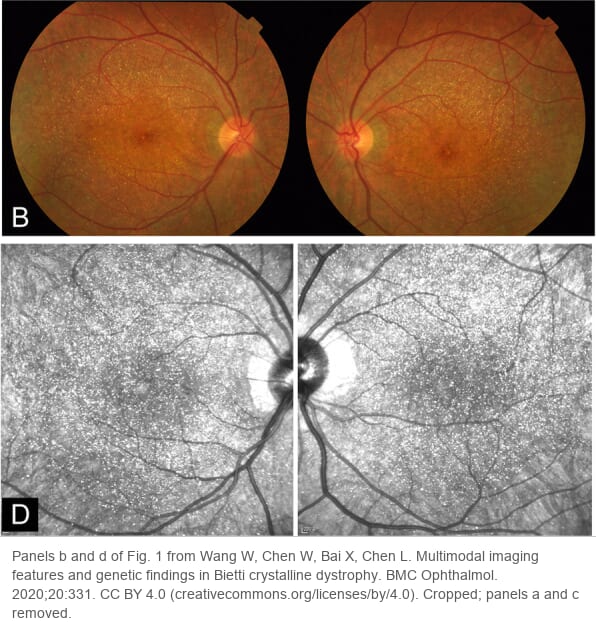
The key sign is numerous fine, glittering yellow-white crystals scattered over the posterior pole. This is where the name "crystalline" comes from. Crystals are easier to see on near-infrared reflectance than on a color photograph[2].
Fundus autofluorescence
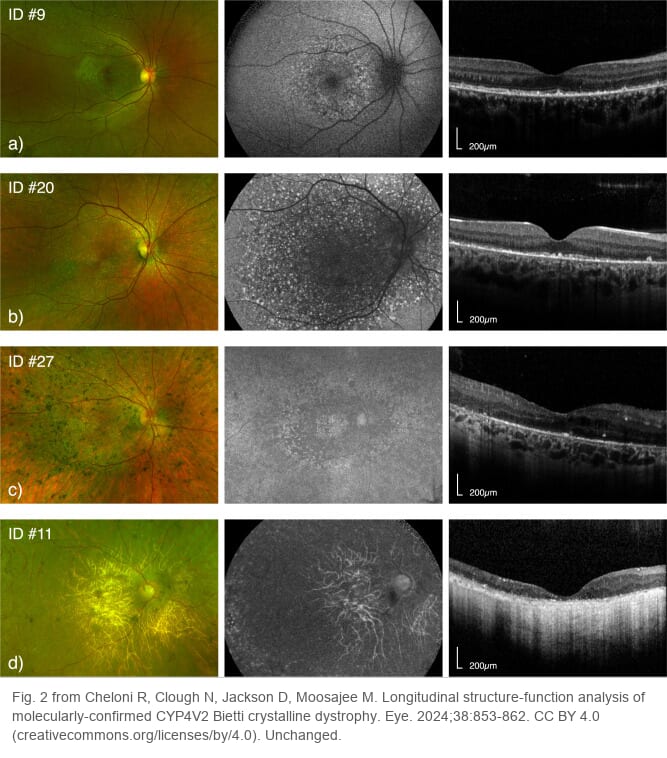
Autofluorescence shows patchy dark (hypoautofluorescent) areas, where the RPE has died. The crystals tend to sit between or around these atrophic patches. As the patches grow and merge, the crystals become smaller and fewer.
ERG
The full-field ERG records the response of the whole retina. Rod and cone responses are both reduced. Early on the ERG can still be normal, and in advanced cases it can no longer be recorded[3].
Telling it from retinitis pigmentosa
Before you settle on RP, check these points.
- The retinal vessels are usually normal. They narrow only in very advanced cases[3].
- Crystals at the corneal limbus are seen in about a third of patients[4]. Look at the limbus with the slit lamp.
- Genetic testing for CYP4V2 helps confirm the diagnosis.
CYP4V2, lipids and treatment
CYP4V2 codes for an enzyme involved in fatty acid metabolism. The crystals are thought to be deposits of abnormal lipid products, and changes in blood fatty acid metabolism have been reported in patients. There is no proven treatment yet[5].
Summary
- Bietti crystalline dystrophy is autosomal recessive (CYP4V2) and more common in East Asia.
- Night blindness starts in the 20s or 30s. Fine glittering crystals lie over the posterior pole, and near-infrared images show them best.
- Autofluorescence shows patchy dark areas of RPE atrophy. Rod and cone ERG responses are usually both reduced.
- Late cases lose their crystals and look like RP. Retinal vessels that are usually normal, limbal crystals and genetic testing tell them apart.
References
[1] Salmon JF. Kanski’s Clinical Ophthalmology: A Systematic Approach. 10th ed. Elsevier; 2025. Chapter 15, Inherited retinal degenerations: Bietti crystalline corneoretinal dystrophy.
[2] Wang W, Chen W, Bai X, Chen L. Multimodal imaging features and genetic findings in Bietti crystalline dystrophy. BMC Ophthalmol. 2020;20(1):331. PMID 32799831.
[3] Rossi S, Testa F, Li A, Yaylacioğlu F, Gesualdo C, Hejtmancik JF, Simonelli F. Clinical and genetic features in Italian Bietti crystalline dystrophy patients. Br J Ophthalmol. 2013;97(2):174–179. PMID 23221965.
[4] Cheloni R, Clough N, Jackson D, Moosajee M. Longitudinal structure-function analysis of molecularly-confirmed CYP4V2 Bietti crystalline dystrophy. Eye (Lond). 2024;38(5):853–862. PMID 37898718.
[5] Ikeda F. Crystalline retinal dystrophy (in Japanese). In: Konnichi no Ganshikkan Chiryo Shishin [Today’s Treatment of Eye Diseases]. 3rd ed. Igaku-Shoin; 2016:533–534.
Figure credits
Fundus and near-infrared images: panels b and d of Fig. 1 from Wang W, Chen W, Bai X, Chen L, BMC Ophthalmol 2020;20:331, licensed under CC BY 4.0. Cropped; panels a and c removed.
Four-patient figure: Fig. 2 from Cheloni R, Clough N, Jackson D, Moosajee M, Eye 2024;38:853–862, licensed under CC BY 4.0. Unchanged.