Coats Disease: Fundus Findings, Fluorescein Angiography and Treatment
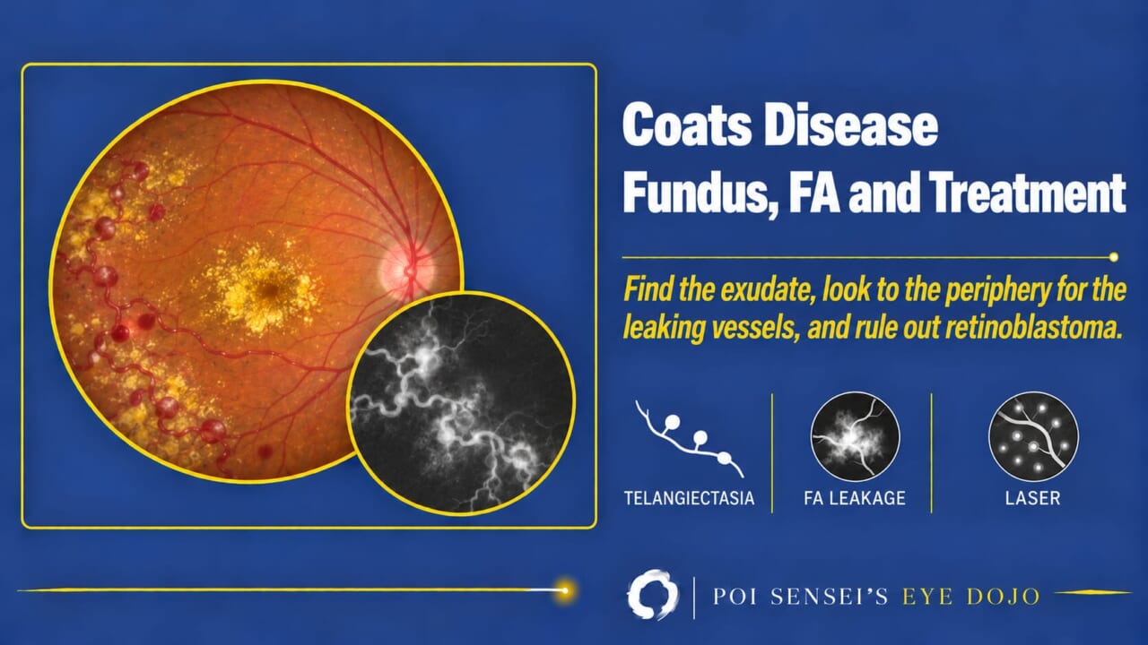
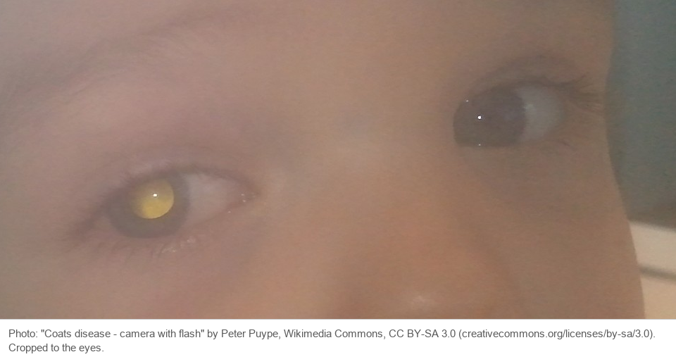
A young boy has a poor red reflex in one eye. The fundus shows yellow exudate, and somewhere in the periphery there are dilated, leaking vessels. This article shows how to recognize Coats disease on the fundus and on fluorescein angiography, and how it is treated.
- In a child with a white pupil, think of retinoblastoma first.
- Who: usually a young boy, and only one eye.
- Find the yellow exudate. It often sits in the macula.
- Look away from the exudate, toward the temporal periphery, for dilated vessels and aneurysms.
- On fluorescein angiography, the abnormal vessels leak and the retina around them is nonperfused.
What is Coats disease?
Coats disease is an idiopathic disease of the retinal vessels. The vessels dilate (telangiectasia) and leak, and exudate builds up in and under the retina.
It is not inherited. Most patients are boys, and it is almost always in one eye. Most present before age 16, but adults can have it too[1].
Two points matter most:
- In young children, it is a cause of leukocoria (a white pupil).
- In severe cases, it leads to exudative retinal detachment and neovascular glaucoma.
Fundus findings
Abnormal vessels
The retinal vessels are dilated and tortuous, with telangiectasia and aneurysms, some shaped like light bulbs. They are often in the temporal periphery.
Exudate
Fluid leaking from the vessels makes the retina swell and leaves yellow lipid (hard exudate) in and under it. When the fluid collects under the retina, it causes an exudative retinal detachment.
The exudate often collects far from the leaking vessels, especially in the macula. So when you see a lot of yellow exudate in one eye of a young patient, do not stop at the macula. Look out to the periphery and find the vessels that are leaking.
Fluorescein angiography
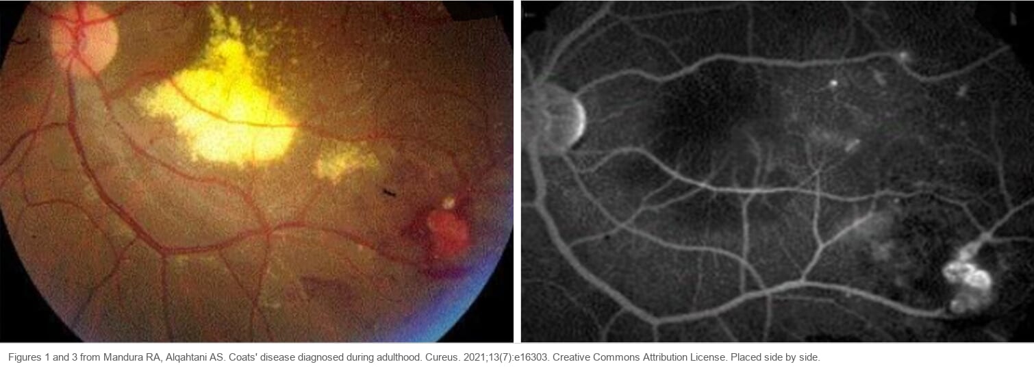
On fluorescein angiography, the telangiectatic vessels and aneurysms light up early and leak late. The retina around them is nonperfused (no blood flow)[2].
Unlike a branch retinal vein occlusion, the nonperfusion is not limited to the territory of one vein. It spreads over areas supplied by several vessels.
Similar to MacTel type 1
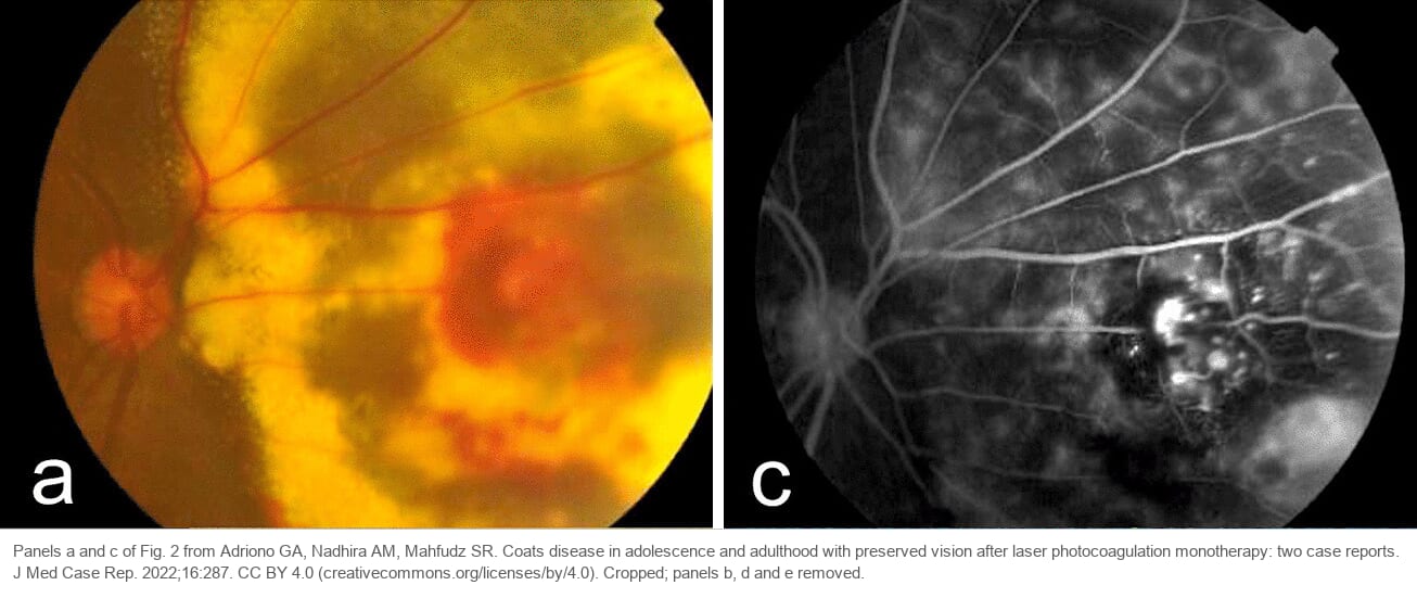
One eye, hard exudates, telangiectasia and aneurysms: macular telangiectasia type 1 shows the same findings, centered on the macula. The two are thought to lie on the same spectrum, with MacTel type 1 at the milder end[1].
Treatment
The aim is to close the leaking vessels so that the exudate can be absorbed.
- Laser photocoagulation is the main treatment. Treat the abnormal leaking vessels directly.
- Cryotherapy is used when extensive subretinal fluid makes laser difficult.
- Anti-vascular endothelial growth factor (anti-VEGF) injections may be added to laser. Evidence is still limited, and long-term safety in children is not known.
- Vitrectomy is for severe eyes with a total exudative detachment or traction (scar tissue pulling on the retina).
Treatment often has to be repeated. The exudate often takes months to clear, and new leaking vessels can appear years later, so children need long follow-up[2].
Summary
- Coats disease: dilated, leaking retinal vessels with exudate, usually in one eye of a young boy.
- In a child with leukocoria, rule out retinoblastoma.
- The exudate collects in the macula, far from the leak. Look to the periphery for the vessels.
- On fluorescein angiography: leaking telangiectasia and nonperfusion over several vascular territories.
- Treat the leaking vessels with laser, or cryotherapy when there is a lot of subretinal fluid, and follow for years.
Figure credits
Leukocoria photograph: "Coats disease – camera with flash" by Peter Puype, Wikimedia Commons, licensed under CC BY-SA 3.0. Cropped to the eyes. The cropped image is shared under the same license.
Fundus photograph and angiogram of adult Coats disease: Figures 1 and 3 from Mandura RA, Alqahtani AS, Cureus 2021;13(7):e16303, distributed under the Creative Commons Attribution License. Placed side by side.
Fluorescein angiography: panels a and c of Fig. 2 from Adriono GA, Nadhira AM, Mahfudz SR, J Med Case Rep 2022;16:287, licensed under CC BY 4.0. Cropped; panels b, d and e removed.
References
[1] Salmon JF. Kanski’s Clinical Ophthalmology: A Systematic Approach. 10th ed. Elsevier; 2025. Chapter 13, Retinal vascular disease: Coats disease.
[2] Yanoff M, Duker JS, eds. Ophthalmology. 5th ed. Elsevier; 2019. Chapter 6.25, Coats’ disease and retinal telangiectasia.