Ethambutol Optic Neuropathy: Signs, Screening and What to Do
Ethambutol is a mainstay drug for tuberculosis and for nontuberculous mycobacterial (NTM) lung disease, such as Mycobacterium avium complex (MAC). Its main eye side effect is ethambutol optic neuropathy: a slow, painless loss of vision in both eyes.
This article covers when it appears, what the eyes show, why the field can look like a chiasmal lesion, and how to screen and manage it.
When it appears
Ethambutol optic neuropathy most often appears 3 to 6 months after starting the drug. It can also appear within the first month, or after more than a year.
The risk depends on the dose and the duration. It is about 1–2.5% at 15 mg/kg a day, 5–6% at 25 mg/kg, and up to 18% at 35 mg/kg[1].
Other risk factors are:
- Kidney disease: ethambutol is cleared by the kidneys, so it builds up
- Older age and low body weight
- Diabetes and hypertension
A dose of 15 mg/kg a day or less makes toxicity much less likely, but it can still happen at that dose. Every patient on ethambutol needs to be watched.
Symptoms and findings
Patients notice blurred central vision in both eyes, often with trouble reading or telling colors apart.
The drug reaches both optic nerves, so both eyes are usually affected to a similar degree. The relative afferent pupillary defect (RAPD) compares the two eyes, so it is often absent. An RAPD can appear when one eye is affected first or more severely.
The typical field defect is a central scotoma, or a cecocentral one that runs from the center to the blind spot. The field can also show a bitemporal defect, a loss of the outer half of the field in both eyes, so it has to be told apart from a chiasmal lesion such as a pituitary tumor.
The critical flicker fusion frequency (CFF) drops, and color vision is affected. Red-green defects are the most common, but blue-yellow defects can come first. Color vision can change before acuity does. A normal Ishihara test does not rule out ethambutol toxicity.
The optic disc
Early on, the fundus is often normal. Some patients have hyperemic, slightly swollen discs, sometimes with small hemorrhages around them.
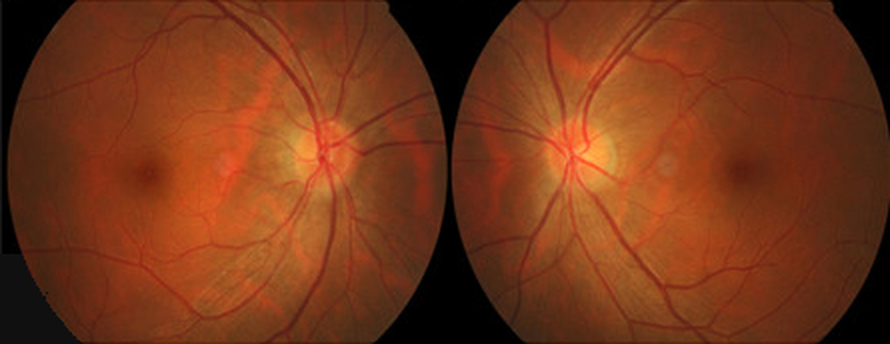
Both discs are red, and their margins are blurred.
Later, optic atrophy sets in, and the disc turns pale, first on the temporal side.
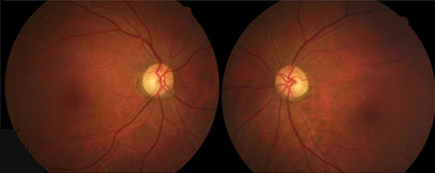
Both discs are pale on the temporal side.
Why it happens
Ethambutol binds metals such as iron, copper and zinc. Zinc deficiency is thought to impair mitochondrial function in the nerve.
One explanation for the bitemporal defect is that the chiasm holds little zinc. When zinc runs short, the chiasm is thought to be hit first, and the crossing fibers carry the temporal fields of both eyes.
Treatment
The treatment is to stop ethambutol. Nothing works better. Contact the prescribing physician right away so that they can switch the regimen.
After that, you can add:
- Vitamin B12 by mouth
- Zinc by mouth if the blood level is low (check serum zinc)
Their benefit is not proven.
Recovery often begins 6 to 8 weeks after stopping, but it can take about a year.
Many patients improve, but recovery is often incomplete. If the drug was stopped late and acuity has already fallen to 20/200 or worse, the outlook is poor[3]. That is why early detection matters.
Screening
In Japan, three societies (tuberculosis, ophthalmology and neuro-ophthalmology) issued a joint statement in 2022[4]:
- Before treatment, the patient brings a referral letter from the prescribing physician. Check acuity, the anterior segment and the fundus.
- During treatment, if there are no symptoms, examine the patient every 1 to 3 months. Check corrected acuity and the visual field, and add the CFF test when needed. Color vision testing and an Amsler grid help detect it early.
- At home, every morning, the patient covers one eye at a time and reads newspaper or phone text at a fixed distance. If it is harder to read than the day before, the patient stops the drug and sees an ophthalmologist.
Before treatment, also record any existing optic nerve disease, such as glaucoma. It makes later changes easier to judge[3].
The US label also asks for monthly acuity checks, one eye at a time, when the dose is above 15 mg/kg a day[5].
Summary
- In bilateral vision loss on TB or NTM treatment, ask about ethambutol, even if it started a year ago.
- Both eyes are usually affected to a similar degree, so the RAPD is often absent.
- The field shows a central scotoma, and sometimes a bitemporal defect.
- Stop the drug first, and warn the patient that vision can worsen for about 3 months.
- Check before treatment and every 1 to 3 months during it, and have patients check each eye every morning.
References
[1] Saxena R, Singh D, Phuljhele S, et al. Ethambutol toxicity: Expert panel consensus for the primary prevention, diagnosis and management of ethambutol-induced optic neuropathy. Indian J Ophthalmol. 2021;69(12):3734–3739. PMID 34827033.
[2] Special issue: Better care for optic neuropathies. MB OCULISTA. 2014;(13):68. Zen-Nihon Byoin Shuppankai. (In Japanese.)
[3] Ministry of Health, Labour and Welfare (Japan). Manual for handling disorders due to adverse drug reactions: retinal and visual pathway disorders. Revised September 2019. https://www.nichigan.or.jp/Portals/0/resources/news/m_484_1.pdf (In Japanese.)
[4] Japanese Society for Tuberculosis and Nontuberculous Mycobacteriosis, Japanese Ophthalmological Society, Japanese Neuro-Ophthalmology Society. Statement on ocular side effects of ethambutol. Nippon Ganka Gakkai Zasshi. 2022;126(3):109–110. (In Japanese.)
[5] Ethambutol hydrochloride tablets, USP. Prescribing information. DailyMed, US National Library of Medicine. Updated November 8, 2023. https://dailymed.nlm.nih.gov/dailymed/drugInfo.cfm?setid=08decdf3-a397-42e7-8ac3-e489c0cc7085

Which retinal layer makes which wave, the three abnormal patterns, and the dark-adaptation trap to check before you report “negative” — on one page, by email. One email when I publish something new, about once a month. Unsubscribe anytime.