Leber Hereditary Optic Neuropathy (LHON): Signs, Genetics and Treatment
A young man notices a blur in the center of vision in one eye. A few weeks later, the other eye loses central vision too. His vision is poor, yet his pupils still react surprisingly well. That picture should make you think of Leber hereditary optic neuropathy (LHON).
This article covers how LHON presents, what the disc looks like, how it is inherited, which mutations to test for, and what can be done.
How LHON presents
LHON typically affects young men around age 20. Women can be affected too, but far less often.
It starts with subacute, painless loss of central vision in one eye. The other eye follows weeks to months later, usually within about two months and almost always within a year[1].
The visual field shows a central scotoma, or a cecocentral one that runs from the center to the blind spot. Acuity often falls to 20/200 or worse, down to counting fingers, but loss of all light perception is rare.
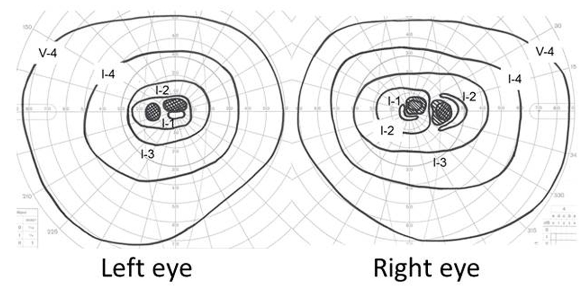
This patient, a 40-year-old woman with vision loss in both eyes, had a small, dense central scotoma in each eye, while the peripheral field was full.
The pupils react better than you would expect from the vision. Even when one eye is much worse, the relative afferent pupillary defect (RAPD) on the swinging flashlight test can be weak or absent. The critical flicker fusion frequency is also often better preserved than the acuity.
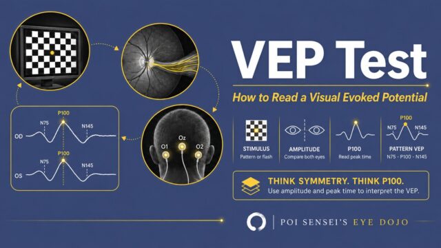
In my own practice, a child was referred with suspected functional (psychogenic) visual loss. The fundus looked normal, the pupils reacted normally, and the field was not reproducible. The VEP, however, showed a delayed P100 in one eye. That pointed to an optic nerve problem, and it led us to the diagnosis of LHON.
The optic disc
In the acute stage, the disc looks red and hyperemic, like ripe fruit. Around it, look for fine, dilated vessels (telangiectatic microangiopathy) and tortuous vessels.
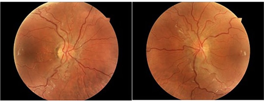
Both discs are red, their margins are blurred by the swollen nerve fiber layer, and the vessels around them are tortuous.
The disc looks swollen because the nerve fiber layer around it is thickened. But this is not true disc edema: fluorescein angiography shows no leakage. This is where LHON differs from optic neuritis with a swollen disc.
OCT shows the thickened nerve fiber layer even when the disc margins look sharp. Within weeks, the disc turns pale, first on the temporal side, and the nerve fiber layer thins.
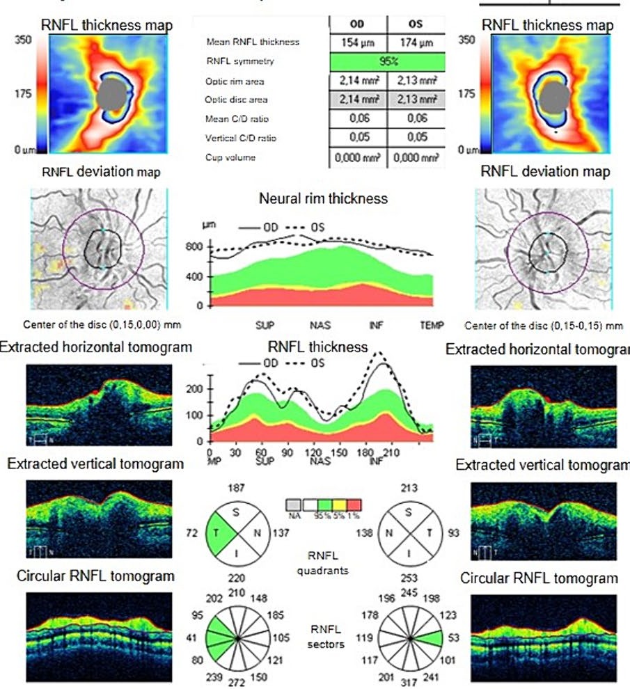
In this patient, the mean peripapillary nerve fiber layer was 154 µm thick in the right eye and 174 µm in the left, and most sectors were white, meaning thicker than the normal range.
About 20% of patients with LHON have a normal-looking disc, and some are labeled as having functional visual loss[1].
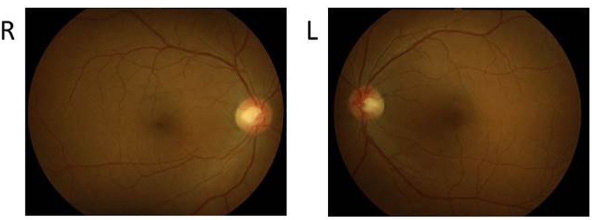
The woman whose visual fields appear above had a fundus that looked normal at the first visit, and fluorescein angiography was normal too.
Inheritance
LHON follows mitochondrial (maternal) inheritance. Mitochondrial DNA comes only from the mother, so a mother who carries the mutation passes it to all her children, sons and daughters alike. A man does not pass it on.
Not every carrier loses vision. About 50% of male carriers and 10% of female carriers do[1].
Because so many of the men in the family are at risk, when I diagnose LHON I explain the disease and its inheritance to the family, together with genetic counseling.
The three main mutations
Three mitochondrial DNA point mutations account for about 90–95% of LHON[1]:
- m.11778G>A
- m.3460G>A
- m.14484T>C
Most genetic tests check these three together. The 11778 mutation is the most common in Northern Europe, Australia and East Asia.
The mutation also tells you the outlook. Recovery is least likely with 11778 and most likely with 14484, with 3460 in between. Even with 11778, about 1 in 10 adults later regain some useful vision[2].
A negative result for all three does not rule out LHON. Rarer mutations also cause it.
Triggers and the heart
Smoking is the clearest trigger. In a study of about 400 carriers, smokers were more likely to lose vision, and heavy smokers more so than light smokers. Heavy drinking showed a weaker trend in the same direction. Head trauma, acute illness and some drugs have also been reported as triggers[1].
So I advise patients and carriers in the family to stop smoking and keep alcohol low.
Heart rhythm problems are more common in LHON carriers, so it is worth asking a cardiologist to check.
Treatment
No treatment reliably restores vision yet.
Idebenone, a synthetic analogue of coenzyme Q10, has been approved in the European Union since 2015 for visual impairment in adolescents and adults with LHON[3]. In its main trial (900 mg/day), it did not clearly beat placebo on the main measure of vision. In a further analysis of the same trial, patients whose two eyes still differed in acuity seemed to benefit[4].
Gene therapy for the 11778 mutation, injected into the vitreous, has also been tested. In one trial, vision improved over two years in both the treated eye and the other eye, which had a sham (mock) injection, so the treatment effect remains unclear[2].
Summary
- A young man loses central vision in one eye, then the other; the pupils react better than the vision suggests.
- The acute disc is red, with telangiectatic vessels and swelling but no leakage; it can also look normal.
- Maternal inheritance: about half of male carriers and a tenth of female carriers lose vision.
- Test for 11778, 3460 and 14484; 11778 has the worst outlook.
- Advise against smoking; idebenone is approved in the European Union.
References
[1] Yu-Wai-Man P, Griffiths PG, Chinnery PF. Mitochondrial optic neuropathies – disease mechanisms and therapeutic strategies. Prog Retin Eye Res. 2011;30(2):81–114. PMID 21112411.
[2] Newman NJ, Yu-Wai-Man P, Carelli V, et al. Efficacy and safety of intravitreal gene therapy for Leber hereditary optic neuropathy treated within 6 months of disease onset. Ophthalmology. 2021;128(5):649–660. PMID 33451738.
[3] European Medicines Agency. Raxone (idebenone). https://www.ema.europa.eu/en/medicines/human/EPAR/raxone
[4] Klopstock T, Yu-Wai-Man P, Dimitriadis K, et al. A randomized placebo-controlled trial of idebenone in Leber’s hereditary optic neuropathy. Brain. 2011;134(Pt 9):2677–2686. PMID 21788663.
Which retinal layer makes which wave, the three abnormal patterns, and the dark-adaptation trap to check before you report “negative” — on one page, by email. One email when I publish something new, about once a month. Unsubscribe anytime.