Horner Syndrome: Confirm It, Then Find the Lesion
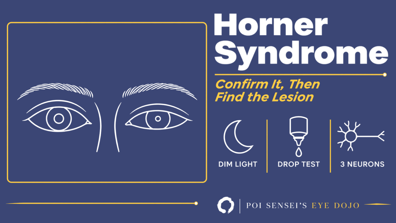
You notice that one pupil is a little smaller and the lid above it sits a little lower. It may be Horner syndrome, a lesion of the sympathetic pathway to the eye. With Horner syndrome you have two jobs: first confirm it, then find where along the pathway the damage is.
What the patient looks like
Horner syndrome has three classic signs on the affected side:
- Miosis. The iris dilator muscle loses its sympathetic supply, so the pupil is small.
- A mild ptosis, usually 1 to 2 mm. The sympathetic supply to Müller’s muscle in the upper lid is lost.
- Reduced sweating on the same side of the face.
A useful clue is the lower lid. In other kinds of ptosis only the upper lid droops, but in Horner syndrome the lower lid also sits slightly higher, because it has a small sympathetic muscle too. The palpebral fissure narrows from both sides, and the eye can look sunken.
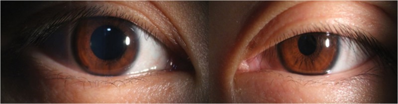
The photo is taken in dim light. Compare the left eye (on the right of the photo) with the right: the upper lid is lower and the pupil is smaller.
The sweating sign is less reliable. The sweat fibers to the face run with the external carotid artery, so the face sweats less only when the lesion is in the first or second neuron, before the sweat fibers branch off at the superior cervical ganglion. Patients sometimes think the normal side is sweating too much.
The pupil: worse in the dark
The sphincter is normal, so the light reaction and the near reaction are normal. What fails is dilation. In bright light both pupils are small and the difference is hard to see. In the dark the normal pupil opens widely and the Horner pupil does not, so the anisocoria becomes obvious.
The Horner pupil also dilates slowly. The anisocoria is largest 4 to 5 seconds after the lights go off. After 10 to 20 seconds the Horner pupil has dilated more, and the difference becomes smaller. This is dilation lag. To see it, turn off the room lights and watch both pupils with a dim light from below. For the full approach to unequal pupils, see Anisocoria.
Three neurons, and what damages each
The sympathetic pathway to the eye has three neurons, and a lesion of any of them causes Horner syndrome.
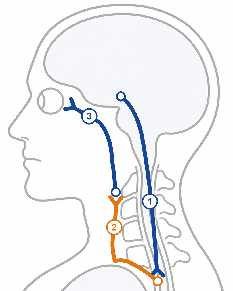
The numbers mark the first, second and third neurons described below.
- First neuron (central): from the hypothalamus down the brainstem to the ciliospinal center in the spinal cord at C8 to T2. A typical cause is a lateral medullary (Wallenberg) infarct. Other brainstem strokes, tumors, demyelination and syringomyelia also do it.
- Second neuron (preganglionic): from the spinal cord over the apex of the lung and up the neck to the superior cervical ganglion. Causes include a tumor at the lung apex (Pancoast tumor), enlarged lymph nodes or a thyroid mass in the neck, an aortic or carotid aneurysm, neck trauma and neck surgery.
- Third neuron (postganglionic): from the superior cervical ganglion up along the internal carotid artery into the cavernous sinus, then to the eye. Causes include internal carotid artery dissection, an aneurysm, a cavernous sinus mass and cluster headache.
So examine the cranial nerves, grip strength (the T1 root) and the neck, including its lymph nodes. A Horner syndrome with a palsy of the third, fourth or sixth nerve points to the cavernous sinus; an internal carotid aneurysm or an internal carotid dissection is one cause to exclude.
Confirm it with drops
About one in five people have a physiological anisocoria, and it too can be a little larger in dim light. It has no dilation lag and no ptosis. When the signs are not clear, drops help to confirm it.
Apraclonidine 0.5% or 1% is the usual test now. Put one drop in both eyes and look again at 30 minutes and, if nothing has changed, at 45 minutes. After the sympathetic supply is lost, the dilator muscle becomes supersensitive, so the Horner pupil dilates while the normal pupil does not. The anisocoria reverses, and the ptosis often improves too. The test detects about 90% of cases, and a positive result is almost always real.
Two cautions:
- The supersensitivity takes several days to develop, so in a very new Horner syndrome the test can be negative. Textbooks suggest cocaine in that situation (see below).
- In young infants, apraclonidine can cross the immature blood-brain barrier, and drowsiness, a slow heart rate and slow breathing have been reported. Avoid it at that age, or watch the baby for a few hours after the drop.
Cocaine 4% works the other way. It blocks the reuptake of noradrenaline at the nerve ending, so a normal pupil dilates. In Horner syndrome no noradrenaline is being released, so the Horner pupil stays small. After cocaine, an anisocoria of 0.8 mm or more in dim light makes Horner syndrome highly likely.
Textbooks still keep cocaine for very new cases, for children, and for an equivocal apraclonidine test when suspicion is high. In practice it is a controlled drug. In Japan it is a narcotic, and cocaine drops are hard to obtain. In the UK they are still used, but they cost more, are harder to get and must be locked away; a London neuro-ophthalmology unit found apraclonidine far more sensitive (93% against 40%) and recommended it as the standard test[1].
For young children some doctors still prefer cocaine, because apraclonidine is debated at that age. The risk of systemic effects from cocaine drops is low, and pressing on the tear ducts after the drop lowers it further. On the other hand, a series of 46 children, 17 of them under 6 months, had no documented adverse effects from 0.5% apraclonidine[2].
Localize it with drops, or with imaging
Drops can also tell a third neuron lesion from a first or second neuron lesion:
| Drop | Normal | 1st or 2nd neuron | 3rd neuron |
|---|---|---|---|
| Phenylephrine 1% | No change | Little or none | Marked |
| Cocaine 4% | Dilates | Little or none | Little or none |
| Tyramine | Dilates | Dilates | None |
Phenylephrine 1% is too weak to dilate a normal pupil. Once a third neuron lesion is about 10 days old, the dilator is supersensitive and dilates strongly. Tyramine and hydroxyamphetamine work by releasing noradrenaline from a living third neuron. If the third neuron is damaged, there is nothing to release, and the pupil stays small.
In practice I have rarely seen these localizing drops used. Hydroxyamphetamine is no longer sold, and the result often does not change the work-up: a carotid lesion can damage either the second or the third neuron. Many clinicians confirm the diagnosis and then image the whole pathway, unless the history or the other signs already show where the lesion is. Still, know these drops: they matter medically, they come up in exams in Japan, and they help you understand the mechanism.
The imaging covers the region from the aortic arch to the circle of Willis: CT or MR angiography, which shows the neck and the carotid arteries, the lung apex, the thyroid and the skull base. Add MRI of the brain if a brainstem stroke is possible.
When it cannot wait
An acute Horner syndrome is an emergency, and urgent imaging looks for a treatable cause. Above all, a new Horner syndrome with pain in the neck, face or head raises carotid dissection.
Here is one of my own patients. A 76-year-old man came with a sudden, severe pain from the top of his head to behind his eye. In the dark his right pupil was 2 mm and his left 5 mm. A right pupil that is smaller in the dark points to a right Horner syndrome. With the sudden pain, we suspected internal carotid artery dissection and performed urgent imaging.
By contrast, if the signs have been present for more than a year and there are no other localizing signs, investigation rarely finds a cause.
Horner syndrome in children
In congenital Horner syndrome the iris on the affected side is lighter, because the iris pigment needs a sympathetic supply during the first year of life. The face on that side may stay pale when the baby cries or feeds, while the other side flushes.
A Horner syndrome in an infant or child, unless surgery near the sympathetic chain explains it, needs a search for neuroblastoma, a treatable tumor. To confirm it in a young child, see the cautions on apraclonidine and cocaine above.
Treatment
Treat the cause. The ptosis itself is mild. If it bothers the patient, apraclonidine drops can lift it for a while, and surgery is an option.
Summary
- A small pupil that lags in the dark, with a mild ptosis, suggests Horner syndrome. Confirm it with apraclonidine.
- A new Horner syndrome with pain is carotid dissection until proven otherwise. Image it urgently.
- In a child with no explained cause, look for neuroblastoma.
References
[1] Bremner F. Apraclonidine is better than cocaine for detection of Horner syndrome. Front Neurol. 2019;10:55. doi:10.3389/fneur.2019.00055.
[2] Maamouri R, Ferchichi M, Houmane Y, Gharbi Z, Cheour M. Neuro-ophthalmological manifestations of Horner’s syndrome: current perspectives. Eye Brain. 2023;15:91-100. doi:10.2147/EB.S389630.
Which retinal layer makes which wave, the three abnormal patterns, and the dark-adaptation trap to check before you report “negative” — on one page, by email. One email when I publish something new, about once a month. Unsubscribe anytime.