Third, Fourth or Sixth Nerve Palsy? How to Work Up Double Vision
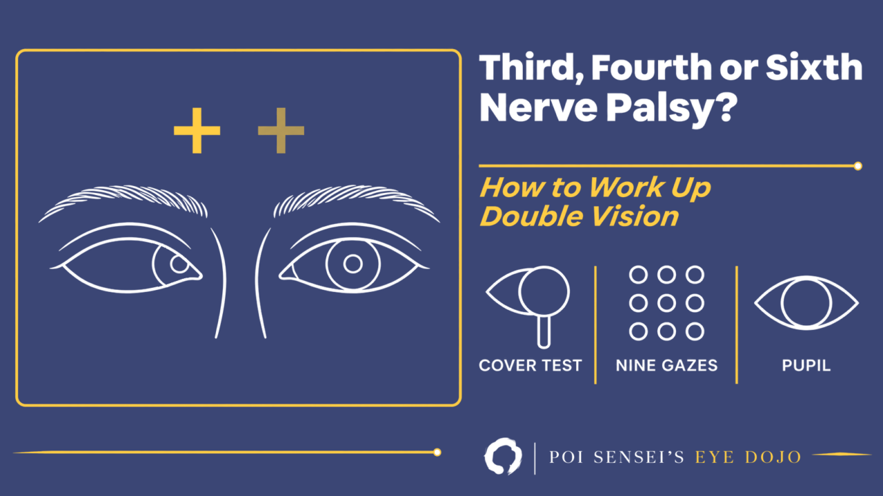
A patient tells you they see double. Before you think of the brain, answer three questions in order. Does the double vision stay when one eye is covered? Is a movement really limited when you test each eye alone? And does the movement problem fit one nerve? The answers tell you who needs urgent imaging.
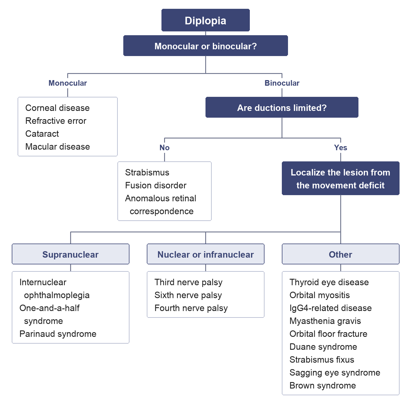
The steps below follow this chart from the top.
Step 1: Does it stay with one eye covered?
Ask the patient to cover each eye in turn. If the second image stays with one eye covered, the diplopia is monocular. Monocular diplopia often comes without any eye movement problem, and the cause is usually in the eye. Dry eye with damage to the corneal epithelium, refractive error and cataract are the common causes; macular disease is another. So examine the ocular surface with fluorescein in every patient with monocular diplopia.
A patient who says "the TV screen looked double" sometimes has a brain CT or MRI before anyone has checked the eye movements. If the diplopia is monocular and the eye movements are full, treating the dry eye may be all that is needed. Do not order brain imaging before you know whether the diplopia is monocular or binocular.
If the diplopia is binocular, ask for details. Is the second image beside the first, above it, or tilted? In which direction of gaze is it worst? Is it worse in the distance or at near? The tips at the end of this article help with the subtle cases.
Step 2: Are ductions really limited?
Test ductions, each eye on its own, as well as versions. In a strabismus, versions can suggest a movement deficit that is not there. In an exotropia, for example, the deviating eye may seem not to adduct when both eyes follow a target to the side. Cover the other eye and test it again: it adducts fully.
A strabismus does not always cause double vision. In a childhood strabismus the image from the deviating eye is usually suppressed. But an old strabismus can break down when the angle changes or the patient is tired, and then new double vision appears.
Step 3: Does the pattern fit one nerve?
When a duction is limited, first ask whether the pattern matches the muscles of one nerve:
- Third (oculomotor) nerve: superior rectus, medial rectus, inferior rectus, inferior oblique, the levator, and the pupil sphincter.
- Fourth (trochlear) nerve: the superior oblique only.
- Sixth (abducens) nerve: the lateral rectus only.
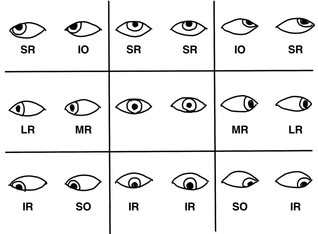
Each cell shows both eyes as you face the patient, looking in one of the nine directions, with the muscle that mainly moves each eye there.
If the pattern fits, think of a lesion of the nucleus or the nerve. If it does not, think of a supranuclear lesion, the neuromuscular junction or the muscle itself. Even a pattern that fits one nerve can come from the neuromuscular junction or the muscle, so keep the mimics further down in mind.
A supranuclear lesion has its own clue. In internuclear ophthalmoplegia, the eye cannot adduct on side gaze, but it can still converge when the lesion is small and anterior, because convergence uses a different pathway. In a third nerve palsy, convergence fails too.
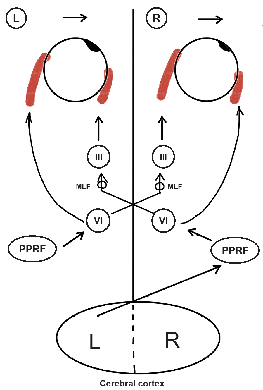
The brain is viewed from above, so the patient’s left is on the left. For a look to the right, the left cortex signals the right PPRF, which drives the right sixth nerve nucleus. Fibers from that nucleus cross and run up the left MLF to the left third nerve nucleus.
Sixth nerve palsy: side-by-side images, worse in the distance
The lateral rectus is weak, so the medial rectus pulls the eye in. There is an esotropia in the primary position and limited abduction on the side of the palsy, with normal adduction. The two images are side by side and uncrossed. The double vision is worse in the distance than at near, and worse when the patient looks toward the side of the palsy. To avoid it, the patient may turn the face toward that side.
In older patients, idiopathic and presumed microvascular palsies are common, up to 60%. Even so, keep a low threshold for imaging, even when the palsy is isolated. Raised intracranial pressure can stretch one or both sixth nerves over the tip of the petrous bone, so a sixth nerve palsy can be a false localizing sign. Test hearing and corneal sensation too: a vestibular schwannoma can damage the sixth nerve; its first symptom is hearing loss, and its first sign is reduced corneal sensation. A bilateral palsy in a child or young adult needs urgent neuroimaging. Any bilateral palsy, a palsy with other neurological signs, and a palsy in a child or younger adult also need thorough investigation. In older patients, ask about symptoms of giant cell arteritis.
Most idiopathic and microvascular palsies recover on their own, up to 90%, over weeks to several months. Until then, a stick-on Fresnel prism or covering one eye relieves the diplopia. In young children, alternate the patching to prevent amblyopia. Surgery is considered only after 6 to 12 months, when recovery has stopped.
Fourth nerve palsy: vertical, tilted, worse looking down
The superior oblique depresses the eye, mostly in adduction, and intorts it. When it is weak, the affected eye sits higher (hypertropia). The hypertropia increases when the patient looks to the opposite side and when they look down, and the eye is extorted. So the double images are vertical, tilted, and worst looking down and in.
Patients describe this in everyday terms. A patient who says the line in the middle of the road looks double while driving is describing diplopia in down-and-in gaze, typical of a fourth nerve palsy. A 68-year-old man with diabetes who says a railway bridge looks tilted is noticing the extorsion. To avoid the double vision, the patient tilts the head to the opposite side and tucks the chin down.
Confirm it with the three-step test, whose third step is the Bielschowsky head tilt test: the hypertropia is worse on gaze to the opposite side and on head tilt to the same side. A Hess chart records the pattern. Note that the side of a fourth nerve lesion and the side of the weak superior oblique are not always the same. The fourth nerve crosses, so a lesion of the nucleus weakens the superior oblique of the opposite eye.
The causes decide what you do next:
- A congenital palsy that has decompensated is common. The double vision feels sudden, but old photographs often show a head tilt, and the vertical fusional range is larger than normal. These patients are usually not aware of the torsion.
- Trauma often damages both fourth nerves. Look for more than 10° of extorsion, a V-pattern esotropia, and a right hypertropia on left gaze with a left hypertropia on right gaze. Do not mistake a bilateral palsy for a unilateral one, especially before surgery.
- Microvascular palsies are common in patients with vascular risk factors. An aneurysm or a tumor is extremely rare.
Routine imaging is not required for an isolated fourth nerve palsy that is not progressing. Consider it if the palsy does not improve. Still, in a prospective study of patients aged 50 or over with an acute isolated fourth or sixth nerve palsy and only vascular risk factors, early MRI found another cause in about 1 in 20, such as a midbrain infarct or a tumor[1]. One pitfall: thyroid eye disease can present with new vertical diplopia and a positive three-step test, so a restriction can look like a fourth nerve palsy.
Congenital decompensated and microvascular palsies often settle without treatment. Persistent double vision can be treated with prisms or surgery.
Third nerve palsy: look at the pupil
A complete third nerve palsy gives a marked ptosis and an eye that is down and out, because the lateral rectus and superior oblique still work. Adduction, elevation and depression are limited. If the parasympathetic fibers are involved, the pupil is dilated and accommodation is weak. A partial palsy gives milder versions of the same signs.
The pupil tells you a lot because of where its fibers run. They lie on the surface of the nerve and get their blood from the pial vessels, while the core of the nerve is supplied by the small vessels inside it.
- A compressive lesion, such as an aneurysm at the junction of the posterior communicating and internal carotid arteries, presses on the surface first, so it usually involves the pupil.
- A microvascular palsy from diabetes or hypertension damages the core of the nerve, so it usually spares the pupil. In diabetes the pupil is spared in about 80%.
This rule is not absolute. A microvascular palsy can involve the pupil, and an aneurysm can spare it at first. The pupil may become involved a few days later as the aneurysm expands. Pupil involvement can also be partial, so even mild pupil signs matter. Pain does not help you decide: marked pain around the eye is common in microvascular palsies too.
What to do:
- An acute third nerve palsy that involves the pupil needs urgent CT angiography to exclude an expanding aneurysm. Be most worried when the pupil is markedly involved while the movement deficit and ptosis are only partial.
- A complete palsy with a normally reacting pupil is much more likely to be microvascular. Microvascular disease is the most common cause, about 40% of cases. Even so, image early: there is consensus on early imaging for third nerve palsies, and in the same prospective study a complete pupil-sparing palsy turned out to be pituitary apoplexy[1]. Assess the vascular risk factors. MRI of the brain and orbits looks for a brainstem, cavernous sinus or orbital apex lesion. For a presumed microvascular palsy, observation is usually appropriate, and most recover over weeks to months.
- Aberrant regeneration, such as the lid lifting when the eye looks down or in, follows trauma or compression. It does not follow a microvascular palsy.
- A third nerve palsy after only a trivial head injury should make you think of an underlying aneurysm or tumor.
Until it recovers, prisms or covering one eye help. Surgery for the deviation and ptosis waits until recovery has stopped, usually at least 6 to 12 months.
When more than one nerve is involved
The third, fourth and sixth nerves and the first division of the fifth run close together in the cavernous sinus and through the superior orbital fissure. A palsy of more than one of them points to the cavernous sinus, the superior orbital fissure or the orbital apex. A Horner syndrome with it suggests the cavernous sinus, because sympathetic fibers run on the carotid artery there. Optic nerve involvement suggests the orbital apex. Causes in the cavernous sinus include an aneurysm, a meningioma, a carotid-cavernous fistula and granulomatous inflammation (Tolosa-Hunt syndrome). Do not hesitate to image and refer these patients.
When the pattern does not fit a nerve
Myasthenia gravis can affect any of the eye muscles, so consider it in any eye movement problem that fits no pattern. It can even look like a pupil-sparing third nerve palsy or an internuclear ophthalmoplegia. The double vision and the ptosis vary, and they are worse at the end of the day. For the ice pack test, place an ice pack on the closed lid for 2 minutes and look for less ptosis; the test is around 75% sensitive but highly specific. Acetylcholine receptor antibodies are found in only 50 to 70% of patients with purely ocular myasthenia, so a negative result does not rule it out.
Miller Fisher syndrome is a rare variant of Guillain-Barré syndrome, and the eye muscles are often the first to be affected. Its triad is ophthalmoplegia, ataxia and areflexia. In about half of patients, all the eye muscles and the pupils are paralyzed, sometimes in both eyes, so the pattern fits no single nerve. It usually follows a viral infection, and anti-GQ1b antibodies are found in most cases. The outlook is good, with full recovery in about 10 weeks on average.
Thyroid eye disease limits movement by restriction, not weakness. A limited elevation may come from a weak superior rectus, or from a stiff inferior rectus that cannot stretch. A forced duction test and MRI of the muscles tell them apart.
Orbital inflammation (idiopathic orbital inflammation and IgG4-related disease) and an orbital floor fracture also limit movement without following one nerve.
Sagging eye syndrome is one of the commonest causes of acquired strabismus in older adults who are not highly myopic. With age, the band that connects the lateral and superior rectus muscles weakens and tears, and the lateral rectus slips down. The patient has a small esotropia in the distance but none at near. If one side sags more, there is a small hypotropia with extorsion on that side. Horizontal ductions are full, though elevation is often limited in both eyes. Ptosis and a deep upper lid sulcus are common clues. MRI shows the torn band and the lowered lateral rectus. Most patients do well with a prism in their distance glasses, because they have no double vision at near; surgery is an option if that is not enough.
Tips for the examination
An obvious movement deficit is easy to recognize, but a mild one often leaves you unsure. Then these help:
- Ask when it happens in daily life. The line in the middle of the road while driving, a tilted bridge, the TV across the room: each points to a direction of gaze.
- Find where it is worst. Check the nine positions of gaze and ask where the double vision is largest.
- Describe the images. Are they side by side, one above the other, or tilted? If side by side, are they uncrossed (an esotropia) or crossed (an exotropia)?
- Think of restriction as well as weakness. A limited movement may come from a weak muscle or from a stiff opposite muscle that cannot stretch. A forced duction test and MRI tell them apart.
Image, refer, but do not over-investigate
Most eye movement disorders need a specialist to finish the work-up, so do not hesitate to image or refer. But do not send every patient with double vision for a brain scan. Diagnose monocular diplopia and decompensated strabismus properly first, and keep urgent imaging for the patients who need it.
Summary
- Cover one eye first: monocular diplopia comes from the eye, not the brain.
- Test ductions, then ask whether the deficit fits one nerve.
- A pupil-involving third nerve palsy needs urgent CT angiography; a normal pupil does not rule out an aneurysm.
- A sixth nerve palsy can be a false localizing sign of raised intracranial pressure.
- If no nerve fits, think of myasthenia gravis, thyroid eye disease and sagging eye syndrome.
References
[1] Tamhankar MA, Biousse V, Ying GS, et al. Isolated third, fourth, and sixth cranial nerve palsies from presumed microvascular versus other causes: a prospective study. Ophthalmology. 2013;120:2264-2269. PMID 23747163.
Which retinal layer makes which wave, the three abnormal patterns, and the dark-adaptation trap to check before you report “negative” — on one page, by email. One email when I publish something new, about once a month. Unsubscribe anytime.