Inherited Retinal Diseases: A Pattern Guide to the Fundus and ERG
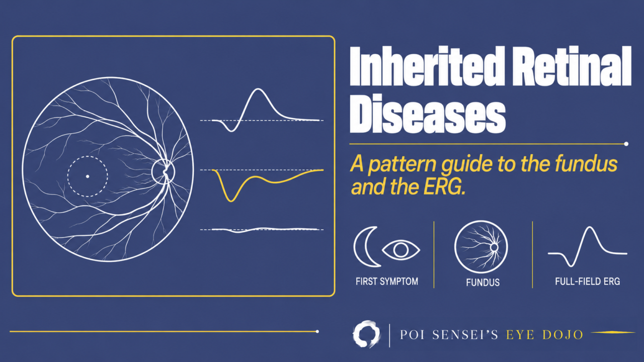
Three questions narrow down an inherited retinal disease: what came first, what the fundus shows and where, and what the full-field electroretinogram (ERG) shows.
- Ask what came first: night blindness, or central vision loss and photophobia.
- Look at what the fundus shows and where: changes mainly at the macula, in the mid-periphery and choroid, white dots, or nothing.
- Record a full-field ERG. Is it normal? Are rods or cones worse? Is the b-wave smaller than the a-wave?
- Finish with autofluorescence, OCT and, for yellow macular lesions, the EOG.
Step 1: Night blindness or central vision first?
Night blindness first means the rod system is failing. Central vision loss, photophobia or poor color vision first means the macula or the cones are failing.
Step 2: What does the fundus show, and where?
Mainly the macula
| Fundus | Think of |
|---|---|
| Atrophy with yellow flecks | Stargardt disease |
| One round yellow lesion | Best disease |
| Many small yellow deposits, also around the disc | Autosomal recessive bestrophinopathy (ARB) |
| A round, sharply bordered patch of atrophy | Central areolar choroidal dystrophy (CACD) |
| Bull’s eye, or near normal | Cone dystrophy |
| Fine radial folds at the fovea in a boy | X-linked retinoschisis (XLRS) |
The mid-periphery and the choroid
| Fundus | Think of |
|---|---|
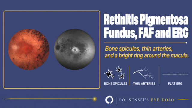
| Bone-spicule pigment, thin arteries | Retinitis pigmentosa (RP) |
| Round, scalloped patches of atrophy that join | Gyrate atrophy |
| Fine glittering crystals | Bietti crystalline dystrophy |
| Choroidal vessels over a pale fundus, macula spared | Choroideremia |
| Round pigment clumps along the arcades | Enhanced S-cone syndrome (ESCS) |
White dots or a golden sheen
| Fundus | Think of |
|---|---|
| Uniform white dots, normal vessels | Fundus albipunctatus |
| White dots with thin vessels | Retinitis punctata albescens |
| A golden sheen that fades in the dark | Oguchi disease |
A normal fundus
A normal fundus does not rule out an inherited retinal disease. Electrophysiology shows where the problem is: the full-field ERG, and the multifocal ERG for the macula.
| Symptoms | Think of |
|---|---|
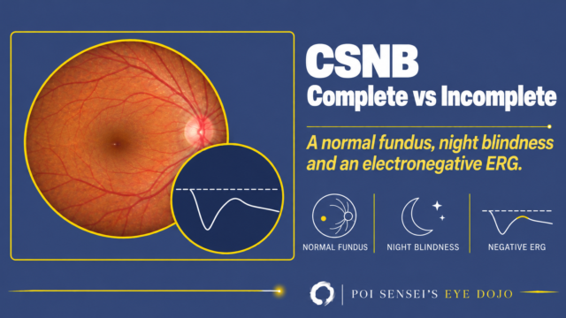
| Night blindness from early childhood that does not get worse | Congenital stationary night blindness (CSNB) |
| No color vision, photophobia and nystagmus from infancy | Achromatopsia |
| Severe visual loss and roving eyes in the first year | Leber congenital amaurosis (LCA) |
| Slow central vision loss in an adult | Occult macular dystrophy |
Step 3: Read the full-field ERG
The full-field ERG adds up the response of the whole retina. In macular disease, a normal full-field ERG means the rest of the retina works: think of a macular dystrophy. A reduced ERG means the disease reaches beyond the macula. The dark-adapted recordings show mainly the rods, and the light-adapted recordings the cones.
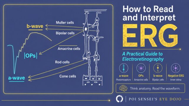
Then look at which cells are failing. The a-wave comes from the photoreceptors and the b-wave mainly from the bipolar cells. If the a-wave is small, the photoreceptors themselves are not working, as in RP. An electronegative ERG is one where the b-wave is smaller than the a-wave. It has three causes: the bipolar cells do not work (CSNB), the signal cannot reach them (XLRS), or the rods are not yet dark-adapted (fundus albipunctatus and Oguchi disease).
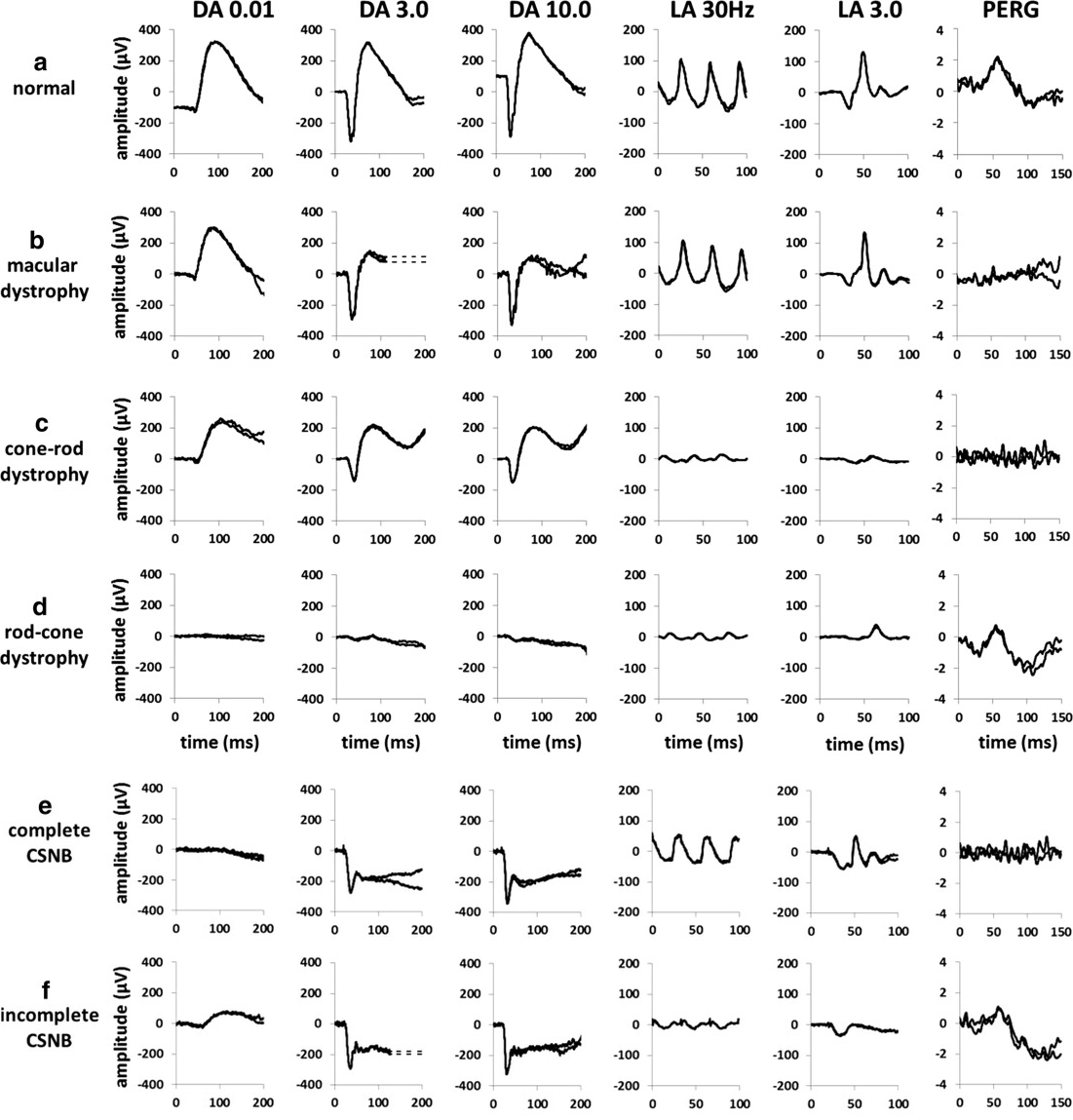
Compare each row with the normal row a:
- b, macular dystrophy: the full-field ERG is normal. Only the PERG is flat.
- c, cone-rod dystrophy: the LA responses are almost flat.
- d, rod-cone dystrophy: the DA responses are flat.
- e, f, CSNB: in DA 3.0 and 10.0, the b-wave is smaller than the a-wave.
| Full-field ERG | Think of |
|---|---|
| Normal | Stargardt (macula only), Best, CACD, occult macular dystrophy |
| Rods and cones reduced, or flat | RP, choroideremia, gyrate atrophy, Bietti, retinitis punctata albescens |
| Cones worse than rods | Cone dystrophy, cone-rod dystrophy |
| Cones absent, rods normal | Achromatopsia |
| Electronegative | CSNB, XLRS |
| Electronegative, improves after hours in the dark | Fundus albipunctatus, Oguchi disease |
| No rod response, the mixed response (DA 3.0) and cone response look alike | ESCS |
| Flat or severely reduced in infancy | LCA |
In fundus albipunctatus and Oguchi disease, the rods take hours to dark-adapt. After the routine 20 to 30 minutes only the cones respond, as in a normal eye with too little dark adaptation. The ERG is electronegative, and unlike CSNB, the a-wave is small too. Record again after a long dark adaptation, and the rod response improves in both.
Step 4: Finish with autofluorescence, OCT and the EOG
Fundus autofluorescence
In Stargardt disease, the atrophy is dark and the flecks are bright. In Best disease, the yellow lesion on the fundus is intensely bright on autofluorescence. In RP, dark patches fill the mid-periphery and a bright ring surrounds the macula. Bietti shows patchy dark areas, and CACD a dark patch with a bright rim.
OCT
In a boy, a retina split into layers in both eyes points to XLRS. In a patient with night blindness, macular cysts that do not leak on fluorescein angiography suggest ESCS. Material or fluid under the retina is seen in Best disease and ARB. Occult macular dystrophy has a normal fundus and a normal full-field ERG. OCT shows a blurred ellipsoid zone (the photoreceptor line on OCT) at the fovea, and the multifocal ERG shows reduced responses at the macula.
EOG for a yellow macular lesion
The electro-oculogram (EOG) tests the function of the retinal pigment epithelium (RPE). In Best disease it is abnormal while the ERG is normal. In ARB it is severely reduced, and the ERG is often abnormal too. An acquired vitelliform lesion in an older adult usually has a normal or only mildly low EOG.
Summary
- Start with the first symptom and where the fundus is abnormal.
- In macular disease, a normal full-field ERG points to a macular dystrophy.
- An electronegative ERG suggests CSNB, XLRS, or the slow dark adaptation of fundus albipunctatus and Oguchi disease.
- Finish with autofluorescence, OCT, the EOG and the multifocal ERG, and rule out acquired causes.
Which retinal layer makes which wave, the three abnormal patterns, and the dark-adaptation trap to check before you report “negative” — on one page, by email. One email when I publish something new, about once a month. Unsubscribe anytime.