Infectious Keratitis: Bacterial, Fungal, Herpetic or Acanthamoeba? From the History to the First Drop
A red, painful eye with a white spot on the cornea. Before any result comes back, you have to decide whether it is an infection, which organism is most likely, what to scrape, and which drops to start.
Three rules run through everything below:
First, is this an infection at all?
An infiltrate with an overlying epithelial defect, a red eye and cells in the anterior chamber points to infection. Anterior chamber cells support infection, but they do not prove it. Herpetic stromal keratitis, an immune reaction, can fill the anterior chamber with cells, and in bacterial keratitis an anterior chamber reaction is common but not always present. Be careful in an eye that is already on a steroid: an infected cornea can look surprisingly quiet, with almost no redness.
Where the infiltrate sits helps too. An infiltrate in the central cornea is more often infectious, and one in the periphery more often non-infectious. That is a tendency, not a rule: a peripheral infiltrate can be infected too.
Three peripheral lesions are commonly mistaken for infection. Marginal (catarrhal) keratitis is not an infection but an immune reaction to staphylococcal antigens from the lid margin, so it comes with blepharitis or meibomitis and sits where the lid margin touches the cornea, separated from the limbus by a clear zone. The epithelial defect is much smaller than the infiltrate, and the anterior chamber shows little or no reaction even when the infiltrate is large. A phlyctenule is a small nodule at the limbus with a leash of vessels running into it. Peripheral ulcerative keratitis and Mooren ulcer are the opposite picture: a peripheral ulcer with little or no infiltrate. When you see one, look for rheumatoid arthritis and other systemic disease.
None of this lets you relax in a contact lens wearer. In a contact lens wearer with a painful red eye, bacterial keratitis has to be ruled out first.
1. Take the history
Most corneal infections come from outside the eye. The organism depends on how it got in and on the patient it got into, so the history narrows the list before you have looked at the cornea.
How fast did it get worse?
Virulent bacteria such as Pseudomonas, pneumococcus and gonococcus can destroy corneal tissue within 24 hours, while nontuberculous mycobacteria and viridans streptococci run a slow course. Fungal keratitis usually starts gradually, with relatively mild pain. A filamentous fungus can then progress fast. Acanthamoeba keratitis progresses very slowly and is often first treated as something else. So the pace is only a starting point.
The amount of pain helps too. Acanthamoeba keratitis is very painful, and herpes simplex keratitis usually hurts only a little.
Also ask about the onset itself. If the patient can name the moment it started, the onset is sudden, and that usually means trauma or an epithelial defect. If it built up over a day or two, the onset is acute.
Contact lenses
Contact lens wear is the most common risk factor for infectious keratitis in high-income countries. The two organisms to worry about are Pseudomonas, which causes more than 60% of contact lens-related keratitis, and Acanthamoeba. The risk is highest with overnight wear, but infection still occurs with careful lens care and with daily disposables.
Reusable lenses add another route: the lens case and solution become contaminated with organisms from the environment, which are then carried onto the eye. So the questions follow that route. Ask what type of lens they wear: daily disposable, reusable soft lenses (two-weekly or monthly), extended-wear, rigid gas-permeable or orthokeratology, or cosmetic lenses bought without a prescription. Ask how they clean them: which solution (multipurpose or hydrogen peroxide), whether they rub and rinse the lens, whether they top off old solution in the case, and how often they replace the case. Ask how many hours a day they wear them, whether they sleep in them, and whether they keep to the replacement schedule. Finally, ask whether the lenses ever meet water: showering, swimming, hot tubs, or rinsing the lens or case in tap water. Look at the other eye too.
Trauma
A patient who first denies any injury often recalls, when asked again, that the pain started after gardening or cutting the grass. People do not count that as trauma. I ask, "Is there anything you think might have caused this?" The answer is often useful.
After trauma, bacteria are the most common cause, but fungi have to be kept in mind. An injury from a tree branch or other plant material is the classic story for filamentous fungi, especially when the keratitis does not respond to antibiotics. Filamentous fungi grow slowly, so a long time can pass between the injury and the first symptoms. An injury can also introduce bacteria and fungi together. A hot metal fragment, such as one thrown off by a grinder, carries a lower risk of infection than a foreign body of plant matter or stone, but any foreign body left in the cornea can become infected. A foreign body can also pass through the cornea into the eye. When you suspect that, consider ultrasonography or CT; CT is the best test for finding a metallic foreign body. MRI is contraindicated when a metallic (especially iron) foreign body may be inside the eye.
Surgery, drugs and past history
Ask about every eye operation, every drop and every tablet. Each answer points to a different organism. A corneal graft, long-term steroid drops or immunosuppression makes you think of yeast (Candida) and of MRSA. After long-term fluoroquinolone drops, streptococci and MRSA are common. Keratitis after laser-assisted in situ keratomileusis (LASIK) makes you think of nontuberculous mycobacteria. Patients with atopic dermatitis get more Staphylococcus aureus and more herpes simplex. Recent shingles on the forehead or nose points to varicella-zoster keratitis. A keratitis that comes back after it had fully settled suggests herpes simplex, and so does an attack after stress or exhaustion. In an older, bed-bound patient with a keratitis that will not heal, think of Pseudomonas, MRSA and fungi.
2. Scrape before the first drop
Telling the organisms apart at the slit lamp is hard, and the signs overlap. Textbooks do describe typical features to some extent, but recognizing them takes experience, and atypical cases are common. So start with a thorough history, make your own prediction from the findings, scrape the cornea, and check your prediction against the smear and culture. Do that every time, and the experience builds up.
Which ulcers to scrape
Scrape any ulcer that is larger than 2 mm, reaches the middle or deep stroma, sits in the visual axis, has lasted a long time, or looks atypical. A small infiltrate, especially one without an epithelial defect and away from the visual axis, may not need a scraping. Most small community-acquired ulcers heal on empirical treatment without a culture. Personally, if I am even slightly unsure, I scrape.
How to scrape
- Instill a topical anesthetic. An unpreserved drop is better, because preservatives can reduce the growth of bacteria. Use proxymetacaine (proparacaine) rather than tetracaine, which is more bacteriostatic.
- Wipe away loose mucus and necrotic tissue from the surface of the ulcer. Look again once it is clean: the true shape of the lesion often shows only then, and it helps to tell bacteria from fungi.
- Scrape the edge of the ulcer, where the organisms are, and the base unless it is thin. Use a blade, a bent needle tip or a spatula. Send the material from the edge for culture in particular: the active organisms are at the edge, while the base may hold dead organisms and debris, which can show on a Gram stain but will not grow. For a suspected fungus, take a firm scrape from the base: hyphae often lie deep in the stroma. When the organisms seem to be deep in the stroma, a small corneal biopsy may be needed.
- Spread a thin smear on one or two glass slides for staining.
- Scrape again for each culture medium and plate the material directly onto the media at the bedside, which gives the best yield. Some laboratories instead take a single scrape in brain-heart infusion broth, or a flocked swab in transport medium, with similar culture-positive rates, so check what your laboratory uses.
For suspected Acanthamoeba, take a wide area of epithelium, including epithelium that looks normal. The infected epithelium is loose and comes off easily. Ask contact lens wearers to bring their lenses, lens case and solution, and send them for culture without cleaning them first.
What the smear tells you on the first day
The Gram stain is the basic stain for bacteria. It tells you gram-positive or gram-negative, cocci or rods, and with the clinical picture that is often enough to choose the first drops. Lancet-shaped gram-positive diplococci, often with a clear halo of capsule, are pneumococcus. Round gram-positive cocci in clusters are Staphylococcus aureus. Pseudomonas gives small gram-negative rods of uneven size, with nothing else distinctive. Moraxella gives large gram-negative rods joined in pairs, the largest bacteria you usually see.
The Giemsa stain shows the inflammatory cells well, but all organisms stain blue, so it cannot tell gram-positive from gram-negative. The cells are a clue in themselves: neutrophils point to bacteria, fungi or Acanthamoeba; mononuclear cells point to a virus; eosinophils point to allergy. Multinucleated epithelial cells suggest herpes simplex or zoster.
Fungi and Acanthamoeba cysts can be seen on a Gram stain too, but Gram and Giemsa stains each pick up only about half of fungal infections. So when you suspect a fungus, add potassium hydroxide (KOH), a quick way to see hyphae, or a fluorescent stain for fungal walls and cysts such as calcofluor white. Under a fluorescence microscope, hyphae, yeast and Acanthamoeba cysts glow, so they are much harder to miss.
Culture and PCR
Plate onto blood agar (most bacteria and fungi), chocolate agar (fastidious bacteria such as Haemophilus, Neisseria and Moraxella), and Sabouraud agar (fungi). For Acanthamoeba, the laboratory uses non-nutrient agar covered with Escherichia coli. Tell the laboratory which organism you suspect: they add selective media, and the yield goes up. If the sample is tiny, tell them which organism to look for first.
Polymerase chain reaction (PCR) is useful for herpes simplex, Acanthamoeba and fungi. For bacteria, PCR of a corneal sample also picks up the normal flora of the ocular surface, so do not diagnose bacterial keratitis from PCR alone.
A culture result needs interpreting. When the same organism is seen on the smear and grown from the corneal scraping, it is almost certainly the cause. An organism grown only from the discharge or the conjunctiva may be a commensal from the lids.
Then start treatment the same day, based on your best guess, without waiting for the culture. Stop contact lens wear in every case. A cycloplegic drop helps the pain and prevents posterior synechiae when the anterior chamber is inflamed.
3. Bacterial keratitis
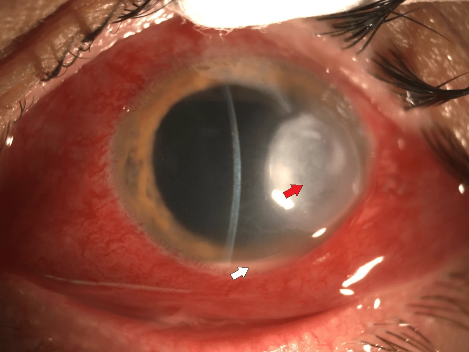
The red arrow points to the ulcer and the white arrow to a small hypopyon.
What it looks like
The typical picture is a white stromal infiltrate with an epithelial defect over it, ciliary injection, and edema of the cornea around the infiltrate. Bacteria digest the stroma, so most infiltrates become ulcers. In moderate to severe cases there is a hypopyon, folds in Descemet membrane, and plaque-like keratic precipitates behind the lesion.
The edema is worth a second look. Recurrent corneal erosion with a little haze can look like early bacterial keratitis, but in an erosion the cornea around the haze is barely swollen. Edema with no opacity at all makes bacteria and fungi unlikely.
The shape of the abscess gives a first hint. A round, localized abscess suggests gram-positive cocci. A ring-shaped abscess suggests gram-negative rods, although Acanthamoeba and fungi can also form a ring.
Pseudomonas is the one to fear. It is usually in a soft contact lens wearer, grows fast with a ring-shaped abscess and a ground-glass cornea around it, and can perforate. Pneumococcus gives a localized abscess after a poke in the eye or with chronic dacryocystitis; the ulcer edge can creep toward the center of the cornea, and it too can go deep and perforate. Staphylococcus gives a localized abscess that is rarely severe. The problem with it is resistance: staphylococci easily become resistant, and MRSA needs a drug such as vancomycin. Moraxella can cause a central keratitis, especially in patients in poor general health. Gonococcus can pass through intact epithelium, so a keratitis can start from a purulent conjunctivitis and perforate quickly. Nontuberculous mycobacteria and Nocardia give faint infiltrates with indistinct borders, after LASIK, trauma or contact lens wear.
Naming the organism from these findings alone takes experience, and atypical cases are common. That is why the Gram stain and culture matter.
What to start
Treatment is intensive topical antibiotics, and the choice depends on the size and site of the ulcer. A small, peripheral ulcer that is not severe can be treated with a fluoroquinolone alone, such as moxifloxacin or levofloxacin. Monotherapy is less toxic to the surface and easier for the patient, and it works about as well as two drugs in most cases.
A large, central or aggressive ulcer, or a smear suggesting streptococci or another specific organism, needs two drugs that cover gram-positive and gram-negative organisms. The standard in the US and the UK is fortified antibiotics: drops made up by the pharmacy at a higher concentration than commercial drops. A typical US pair is cefazolin 5% (vancomycin for MRSA) with tobramycin 1.4%; a typical UK pair is cefuroxime 5% with gentamicin 1.5%.
Give the drops every hour, day and night, for the first 24 to 48 hours, then taper as the eye improves. For a severe ulcer, start with a loading dose, one drop every 5 to 15 minutes, before going hourly. In less severe cases, an ointment can be used at bedtime. Admit patients who cannot manage drops that often, and consider admission for a severe ulcer in an only eye. If the cornea is significantly thinned, a clear shield between drops protects it. Systemic antibiotics are rarely needed. Use them when the infection spreads to the sclera, when perforation is threatened or present, and for gonococcal keratitis, which needs systemic ceftriaxone.
And the steroid?
Do not start a steroid on the first day. If you use one at all, keep it for a culture-proven bacterial ulcer that is already improving on antibiotics, without significant thinning, and not caused by Nocardia. The largest trial, the Steroids for Corneal Ulcers Trial (SCUT), found no overall benefit in final vision. Severe ulcers tended to do better, but every patient in the trial had a positive culture and the steroid was started only after 48 hours of antibiotic. In a later analysis, starting the steroid 2 to 3 days after the antibiotic did better than starting it after 4 days or more. Steroids slow epithelial healing, and Nocardia ulcers did worse on them. The Japanese guideline suggests not adding a steroid.
4. Fungal keratitis
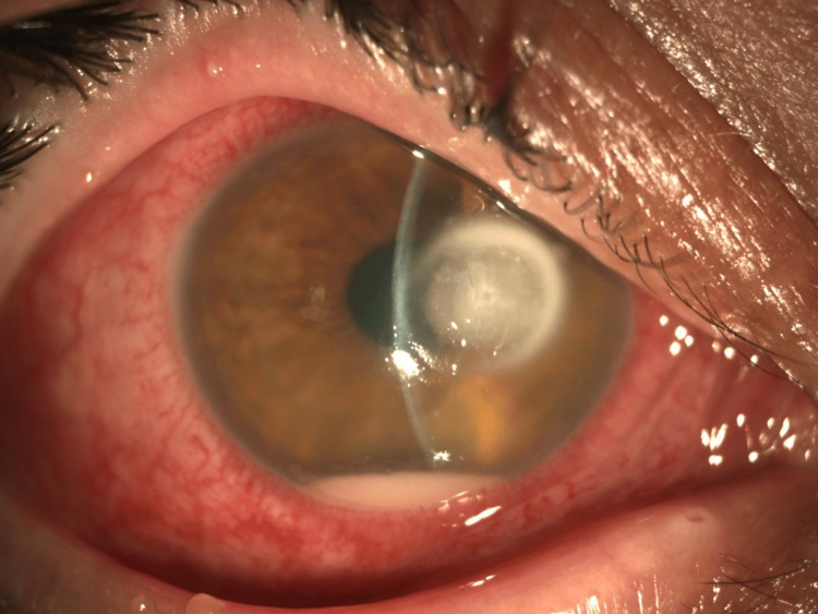
A 4 mm ulcer with a dry look and indistinct, feathery borders, with an endothelial plaque, a hypopyon and fibrin in the anterior chamber.
Two groups, two stories
Fungi split into two groups, and each comes with its own story. Filamentous fungi (Fusarium, Aspergillus) follow trauma, especially from plant material. Yeast (Candida) grows in a compromised eye: a corneal graft, long-term steroid drops or ocular surface disease. Which group you meet also depends on the climate: yeasts cause most fungal keratitis in temperate countries, and filamentous fungi are the most common in the tropics, though they are not rare in cooler regions.
What it looks like
A filamentous fungus gives a gray-white infiltrate with feathery, indistinct margins, often with satellite lesions separate from the main infiltrate. An endothelial plaque, a large disc-shaped deposit on the back of the cornea, is typical, together with a hypopyon. It suggests that hyphae may have reached the deep stroma or the anterior chamber. Early on, the stromal layers can look surprisingly intact even when the infection has gone deep. Fungi tend to go deep, and perforation is more common than with bacteria.
Yeast is harder. It gives a round, well-defined, dense yellow-white infiltrate, often in the superficial stroma, with a strong tendency to melt. That looks just like a gram-positive bacterial keratitis, so here the history and the scraping matter most.
A steroid changes the picture, and a fungal keratitis on steroid drops may not look typical at all. Some filamentous fungi stay in the most superficial stroma, progress very slowly and cause little inflammation, so they can look like a persistent epithelial defect or a corneal deposit.
What to start
When you cannot tell bacteria from fungus, and the keratitis is no worse than moderate, treat it as bacterial first and add antifungal treatment if it does not respond. When the keratitis is severe or a fungus is strongly suspected, start antibacterial and antifungal treatment together.
For filamentous fungi, natamycin 5% is the first choice, especially for Fusarium. It is the only antifungal eye drop approved by the FDA. Voriconazole 1% drops, made up by the pharmacy, are added when natamycin is not working. Do not use voriconazole on its own: in the largest trial, 323 patients in South India, natamycin gave better vision and fewer perforations, mostly because of the Fusarium cases[3]. For yeast, use amphotericin B 0.15% or econazole 1%. Give the antifungal drops every hour for the first 48 hours, then reduce them as the signs allow.
Stop any steroid. Debride the lesion regularly: it removes organisms and lets the drops penetrate, but be careful where the cornea is thin. Systemic antifungals may be given for severe infection, lesions near the limbus and suspected endophthalmitis, but the evidence for them is limited. In a randomized trial of severe filamentous ulcers (MUTT II), adding oral voriconazole to the drops did not reduce perforation or the need for keratoplasty, and side effects such as raised liver enzymes and visual hallucinations were about twice as common[4].
Then be patient. Fungal keratitis responds slowly, and the drops themselves can damage the surface and blur the picture. If it is not getting worse, take that as a response. Judge it by the size of the epithelial defect, the infiltrate and the hypopyon, and keep the same regimen for at least a week before you add or change a drug.
5. Herpes simplex keratitis
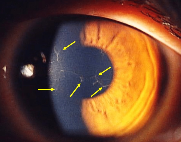
The arrows point to dendrites in the epithelium of an orthokeratology lens wearer.
Herpes simplex keratitis has two main forms, and the mechanism is different, so the treatment is different. The difference that matters is the steroid: epithelial disease gets none, and disciform disease needs one.
Epithelial keratitis: treat the virus
Epithelial keratitis is active virus in the epithelium. The dendritic ulcer branches like the corneal nerves, and the ends of the branches are swollen into terminal bulbs. The edge of the ulcer is slightly raised and gray, so the whole dendrite looks outlined. It has some width, and the epithelium elsewhere is normal. Left untreated or treated with a steroid, a dendrite can widen into a geographic ulcer. A geographic ulcer keeps the outlined edge, and usually a dendritic tail somewhere along it.
Treat the virus. Use acyclovir 3% ointment or ganciclovir 0.15% gel five times a day. Trifluridine 1% drops are an alternative but need up to nine times a day. Oral acyclovir, valacyclovir or famciclovir is also an alternative, and is preferred in children, in immunocompromised patients and when drops are poorly tolerated. Do not use a steroid: it can turn a dendrite into a geographic ulcer.
Disciform keratitis: calm the immune reaction
Disciform keratitis (endotheliitis) is a round zone of central stromal edema, often with epithelial edema over it, and keratic precipitates under it. An immune ring can form around it. An immune reaction is more likely to be involved than in epithelial disease, so treatment needs a steroid together with an antiviral. Pain and redness are often milder than in epithelial disease.
Suppress the inflammation with a topical steroid, and give antiviral cover at the same time, because the steroid can reactivate the virus. Taper slowly over weeks to months; stopping too fast brings it back. A steroid without antiviral cover often works at first and then relapses. Long-term oral acyclovir (400 mg twice a day) reduces recurrences and is worth considering for patients with repeated attacks or a corneal graft. Reduce the dose of oral antivirals in renal impairment.
Corneal sensation is reduced in herpetic keratitis. It also falls with age and long-term contact lens wear, so compare the two eyes.
When a branching lesion is not herpes simplex
A branching lesion without terminal bulbs is a pseudodendrite. Pseudodendrites taper at the ends, have no infiltrate in their edges, and are usually thin. Before you call it herpes, think of four other causes.
Varicella-zoster virus gives small, thin lesions, often radiating from one center like a star. They are raised, stain only faintly with fluorescein, and come with shingles in the first division of the trigeminal nerve. A rash on the tip or side of the nose (Hutchinson sign) raises the risk of eye involvement, but zoster without a rash also exists. Oral antivirals should be started as early as possible for the shingles. Stromal zoster keratitis often needs a stronger steroid than herpes simplex does, and undertreatment leaves scarring, synechiae and glaucoma.
Drug toxicity gives a branching crack line, usually horizontal and just below the center of the cornea, with some opacity and dense punctate keratopathy around it, so ask exactly which drops the patient is using. A healing recurrent erosion leaves loose epithelium around the lesion that lifts off the stroma, and severe pain on waking is typical. And Acanthamoeba can give a pseudodendrite, though almost never alone: look for the other early signs in the next section.
A geographic ulcer also has look-alikes. A persistent epithelial defect or a neurotrophic ulcer is usually a smooth oval with a rolled, gray edge, not a map.
6. Acanthamoeba keratitis
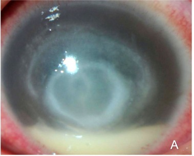
A ring infiltrate in the central stroma, with a hypopyon.
The usual story is a contact lens wearer treated as "herpes" for weeks, often with a steroid, who keeps getting worse. Each stage of Acanthamoeba keratitis has a herpes look-alike: the early pseudodendrite looks like an epithelial herpes dendrite, and the later disc looks like disciform herpes. Keep Acanthamoeba on the list in any contact lens wearer with a pseudodendrite or a disciform lesion.
What it looks like
Most cases are in contact lens wearers, but trauma and contact with contaminated water can also cause it. The key symptom is pain out of proportion to the signs, with strong ciliary injection. The infection starts in the central superficial cornea, spreads slowly outward and only later goes deep, so the signs change with time. The whole point is to suspect it early, so learn the early signs.
In the first few weeks, you see punctate, patchy or linear infiltrates in and under the epithelium, a pseudodendrite, and radial keratoneuritis: linear infiltrates running along the corneal nerves from the limbus toward the center. Radial keratoneuritis is very specific for Acanthamoeba. Limbitis, a swollen and very red limbus with severe pain, can come with it.
Later, as the infiltrates enlarge and join, a ring infiltrate forms in the central stroma, or a disc-shaped infiltrate with edema, sometimes with large keratic precipitates and a hypopyon.
Scrape a wide area of epithelium, as described above, and send the lens case. In vivo confocal microscopy can show Acanthamoeba cysts and fungal hyphae in the living cornea, but it is mostly available in tertiary centers.
What to start
Debride the infected epithelium. It removes organisms, gives you material to examine, and helps the drops penetrate. Repeat it two or three times a week at first.
The main drug is polyhexamethylene biguanide (PHMB) 0.02% or chlorhexidine 0.02%. Both kill the active form and the cysts. Propamidine or hexamidine is often added. Give the drops every hour at first, then taper over months. Relapses are common during the taper. In the European Union, a stronger PHMB 0.08% (Akantior) was approved in 2024 for use on its own. In the trial behind it, it cured about as many patients as PHMB 0.02% with propamidine (85% vs 89%)[5]. Voriconazole 1% drops have been used as an addition or an alternative, but the evidence is limited. Apart from PHMB 0.08% in the EU, these drops are usually not commercially available and have to be made up by a pharmacy, so find out in advance where your nearest supply is.
Avoid steroids, above all before the diagnosis: a steroid given before amoebicidal treatment is a risk factor for a poor outcome. Some specialists add a low dose for persistent inflammation, but only after at least two weeks of amoebicidal treatment, and keep the amoebicide going for weeks after the steroid stops. For pain, use an oral nonsteroidal anti-inflammatory drug. Oral miltefosine is an option when topical treatment fails.
7. The next day and the following week
See the patient again the next day. Improvement shows as less lid swelling and chemosis, a shrinking epithelial defect, a less dense infiltrate, and a quieter anterior chamber. The anterior chamber cells and the height of a hypopyon are useful markers of response.
One pitfall: as the corneal edema clears, the anterior chamber becomes easier to see, and the cells can look more numerous. That is not a sign of worsening.
If the eye is improving, do not change the drops, even if the laboratory reports a resistant organism: topical treatment reaches far higher levels in the cornea than the laboratory tests assume.
If the keratitis is not improving, think of four things:
- The wrong organism. Look at the history and the first findings again, and ask which organisms your drops would not have covered. Bacterial keratitis that does not respond may be fungal or Acanthamoeba. Scrape again if the first culture was negative.
- A mixed infection. Trauma can bring bacteria and fungi together. Herpes simplex keratitis can appear during treatment for bacterial keratitis, especially in patients with atopic dermatitis. A keratitis that was improving and suddenly gets worse suggests a second organism.
- Toxicity from the drops. Frequent drops, especially aminoglycosides and high concentrations, damage the epithelium. Long courses of topical antivirals do the same. A non-healing epithelial defect with punctate erosions around it can be toxicity, and it is easily mistaken for persisting infection. Toxicity can even cause a hypopyon, which makes it look still more like a worsening infection. An epithelial defect that will not heal after herpetic keratitis can also be neurotrophic keratopathy, so test the sensation.
- The drops are not going in. Hourly drops are hard. Ask the patient exactly how they are using them.
If the cornea thins or perforates, lower the intraocular pressure, keep the eye at rest, and keep treating the infection. A small perforation with the infection under control can be closed with a bandage contact lens, or tissue glue under a bandage lens. A larger perforation, or infection that keeps spreading despite treatment, needs a therapeutic keratoplasty. If possible, a keratoplasty for vision is done after the infection has settled.
Summary
- Take the history first: speed of onset, contact lenses, trauma, surgery and drugs.
- Make a guess from the cornea, then scrape the edge of the ulcer before the first drop.
- Start treatment the same day: intensive antibiotics or antifungals, an antiviral, or an anti-amoebic drug.
- When you are not sure of the organism, do not start a steroid.
- Review the next day; if it is not improving, think of the organism, a second infection, toxicity and adherence.
References
[1] Infectious Keratitis Guidelines Committee of the Japanese Association for Ocular Infection. Guidelines for the management of infectious keratitis, 3rd edition. Nippon Ganka Gakkai Zasshi. 2023;127:859-895. (In Japanese.)
[2] Infectious Keratitis Guidelines Committee of the Japanese Association for Ocular Infection. Guidelines for the management of infectious keratitis, 2nd edition. Nippon Ganka Gakkai Zasshi. 2013;117:467-509. (In Japanese.)
[3] Prajna NV, Krishnan T, Mascarenhas J, et al. The Mycotic Ulcer Treatment Trial: a randomized trial comparing natamycin vs voriconazole. JAMA Ophthalmol. 2013;131:422-429. PMID 23710492.
[4] Prajna NV, Krishnan T, Rajaraman R, et al. Effect of oral voriconazole on fungal keratitis in the Mycotic Ulcer Treatment Trial II (MUTT II): a randomized clinical trial. JAMA Ophthalmol. 2016;134:1365-1372. PMID 27787540.
[5] European Medicines Agency. Akantior (polihexanide): EPAR. Marketing authorisation 22 August 2024. https://www.ema.europa.eu/en/medicines/human/EPAR/akantior (accessed 7 November 2026).

Which retinal layer makes which wave, the three abnormal patterns, and the dark-adaptation trap to check before you report “negative” — on one page, by email. One email when I publish something new, about once a month. Unsubscribe anytime.