Bacterial, Viral or Allergic Conjunctivitis? How to Tell Them Apart
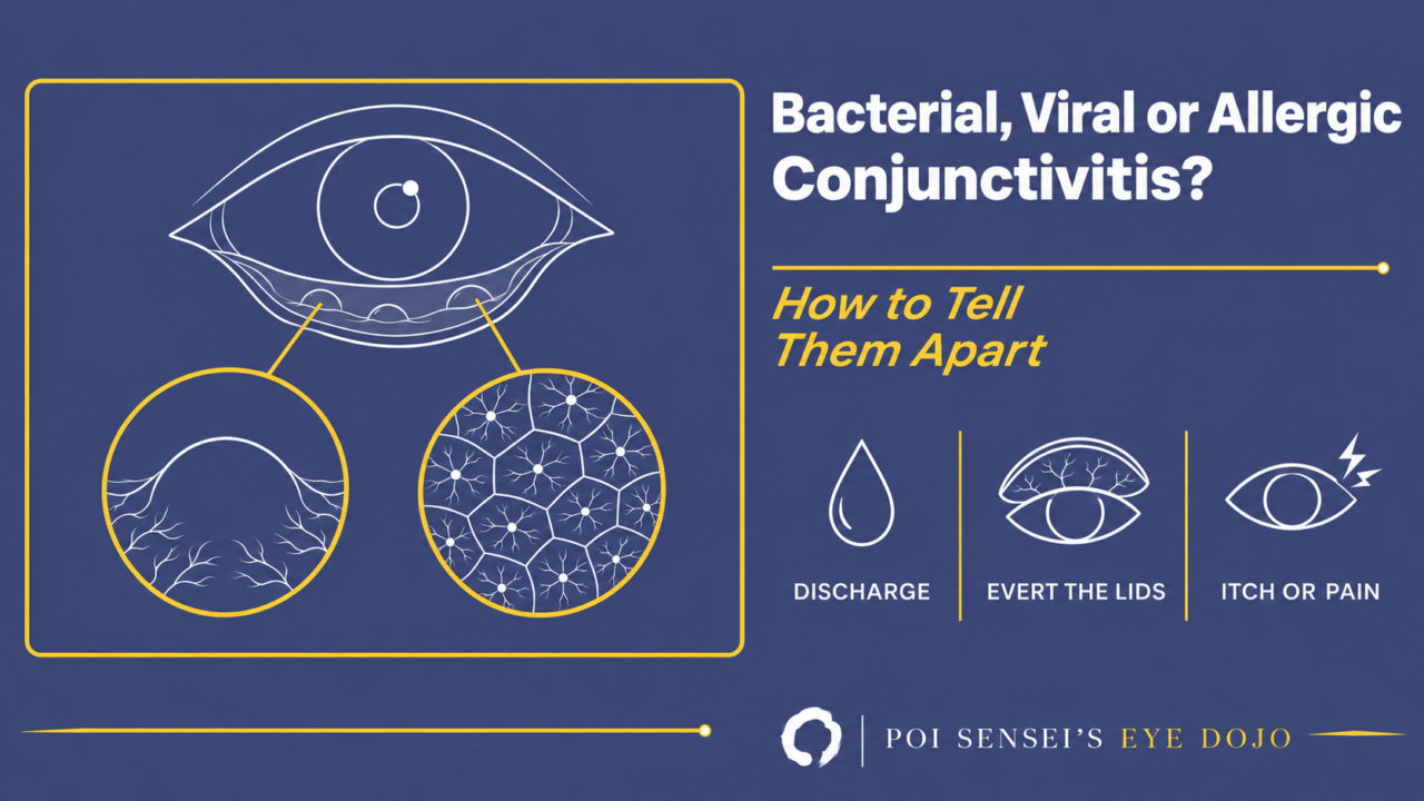
A red eye with discharge is one of the most common reasons to come to an eye clinic, and most of these patients have bacterial, viral or allergic conjunctivitis. The history often seems to give the answer away. A patient who says "This happens every spring" or "My whole family has red eyes" makes it tempting to decide before you look. But deciding from the history alone is how mistakes happen. Look at the rest of the eye first, and then at five things: the discharge, the tarsal conjunctiva, itch or pain, the onset, and who else has a red eye.
First, look past the conjunctiva
Before you sort out the type, look at the rest of the eye.
1. The discharge
Conjunctivitis always comes with some discharge, whatever the cause, and most other causes of a red eye do not. So look at it.
- Bacterial conjunctivitis gives a purulent or mucopurulent, yellow-white discharge. The lids are often stuck together on waking. On the first day it can still be watery, so a watery discharge does not rule bacteria out.
- Viral conjunctivitis gives a watery discharge.
- Allergic conjunctivitis gives a watery discharge in an acute attack. In vernal and atopic keratoconjunctivitis it is often a stringy mucus.
A profuse purulent discharge that is back within seconds after you wipe it away suggests gonococcus. The cornea can perforate within days, so take a swab and treat with systemic ceftriaxone.
2. The tarsal conjunctiva: follicles or papillae
Evert the lids and look for follicles or papillae.
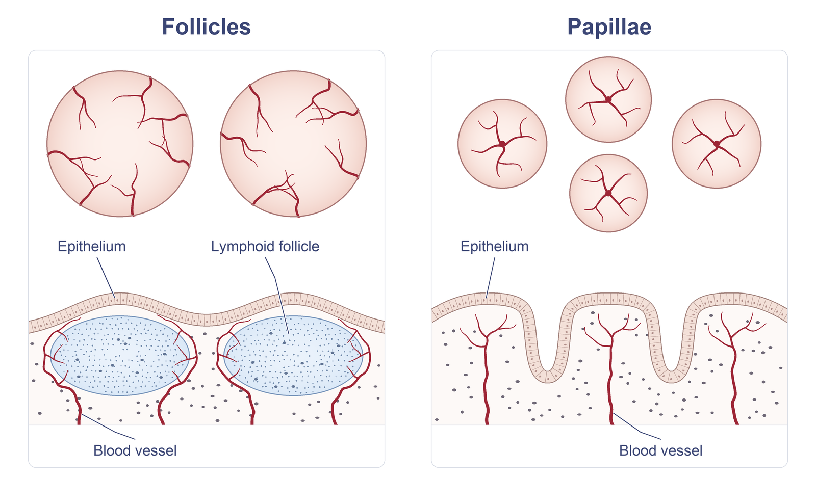
The top row shows single lesions up close, and the bottom row is a cross-section. The red lines are blood vessels: they run across a follicle from its edge, but rise from the center of a papilla.
- A follicle is a mound of lymphoid tissue. Vessels run around it, and there is no vessel at its center. Follicles point to viral conjunctivitis, and also to chlamydia and to a reaction to topical drops.
- A papilla has a vessel at its center. Papillae point to allergic conjunctivitis, but they also appear in bacterial conjunctivitis, blepharitis and contact lens wear.
Two more signs point to adenovirus: small petechial hemorrhages on the conjunctiva and a tender preauricular lymph node. In bacterial conjunctivitis the node is usually not enlarged, except in severe gonococcal infection.
The lids help in one more way. In adenoviral conjunctivitis the tarsal conjunctiva is red too. If the bulbar conjunctiva is very red but the tarsal conjunctiva looks quiet, think of something other than conjunctivitis.
3. Itch or pain
Itch is the hallmark of allergic conjunctivitis. Viral conjunctivitis can itch too, so itch alone does not rule infection out.
Pain, photophobia or a marked foreign body sensation is different. It suggests that the cornea is involved, so look at the cornea before you settle on conjunctivitis.
4. Onset, age, one eye or both, and contacts
- Infectious conjunctivitis starts suddenly. Bacterial conjunctivitis reaches its worst within one to two days. Allergic conjunctivitis comes back in the same season every year, or goes on all year.
- Bacterial conjunctivitis is most common in small children and the elderly.
- Adenovirus usually starts in one eye, and the other eye follows one to a few days later. Allergic conjunctivitis affects both eyes.
- Ask whether anyone at home, at school or at work has a red eye. Adenovirus spreads through families and classrooms.
The typical pattern:
| Bacterial | Viral | Allergic | |
|---|---|---|---|
| Discharge | Purulent | Watery | Watery or mucoid |
| Tarsal | Papillae | Follicles | Papillae |
| Course | Acute | Acute | Recurrent |
Treatment in one line each
- For bacterial conjunctivitis, a topical antibiotic shortens the course. I culture the elderly, patients with atopic dermatitis and any case that does not clear, because MRSA and resistant organisms are more likely in them.
- For adenoviral conjunctivitis, no antiviral works. Give cold compresses and artificial tears, and teach the patient how not to spread it. Do not start a steroid early. Keep it for a pseudomembrane or for subepithelial infiltrates that reduce the vision.
- For allergic conjunctivitis, start a drop that is both an antihistamine and a mast cell stabilizer, such as olopatadine. In severe vernal keratoconjunctivitis, add topical cyclosporine.
The details for each type, including the five types of allergic conjunctivitis, how to read a rapid adenovirus test and how to stop adenovirus spreading in your clinic, are in Chapter 3 of my book, Ophthalmology for Your First Year, Volume 1.
Summary
- Look at the discharge: purulent for bacteria, watery for viruses, watery or mucoid for allergy.
- Evert the lids: follicles favor a virus, papillae favor allergy.
- Itch points to allergy. Pain or photophobia means look at the cornea.
- Do not decide from the history alone. Check the cornea and the anterior chamber in every red eye.

Which retinal layer makes which wave, the three abnormal patterns, and the dark-adaptation trap to check before you report “negative” — on one page, by email. One email when I publish something new, about once a month. Unsubscribe anytime.