Retinopathy of Prematurity (ROP): Zones, Stages, Plus Disease and When to Treat
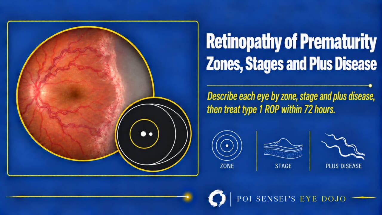
A premature baby’s retina is still growing its blood vessels. Retinopathy of prematurity (ROP) is abnormal growth of these vessels. This article shows how to describe an ROP exam with the international classification (zone, stage and plus disease) and how the three together tell you when to treat.
- Zone: how far back is the disease? Zone I is the most posterior and the most dangerous.
- Stage: what does the edge of the vessels look like? A line, a ridge, or new vessels growing into the vitreous.
- Plus disease: are the vessels around the disc dilated and tortuous?
- Check the three against the treatment criteria. If they are met, treat within 72 hours.
What is ROP?
The retina has no blood vessels until about the fourth month of pregnancy. Then vessels grow out from the optic disc toward the edge of the retina. They reach the nasal edge first, at about 8 months of pregnancy, and the temporal edge last, at or soon after term[1].
This growth is driven by VEGF (vascular endothelial growth factor), a signal that makes vessels grow. The retina ahead of the vessels has no blood supply, so it is short of oxygen and releases VEGF, and the vessels grow toward it. In a baby born early, the growth stops. Later, the avascular retina makes too much VEGF. New vessels then grow abnormally into the vitreous, form fibrous membranes, and can pull the retina off.
Who is screened
Screening criteria and timing differ by country, so follow your local guideline. As a rough guide, babies born at about 30–32 weeks or earlier, or weighing 1500 g or less, are screened. The first exam is usually at 4–7 weeks after birth, and exams are repeated every 1–3 weeks until the vessels reach zone III[1].
Zone
ROP is described with the International Classification of Retinopathy of Prematurity, now in its third edition (ICROP3, 2021). For each eye, record the zone, the stage and plus disease.
The zone tells you how far back the disease is. The zones are circles centered on the optic disc:
- Zone I is a circle with a radius of twice the distance from the disc to the fovea.
- Zone II reaches from the edge of zone I out to the nasal ora serrata.
- Zone III is the crescent of temporal retina left outside zone II.
Find the edge between the vascularized and the avascular retina, and any ROP lesion. The most posterior of these sets the zone. The more posterior the zone, the more immature the retina.
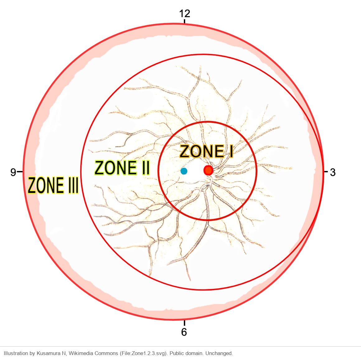
ICROP3 added two terms. Posterior zone II is the band of 2 disc diameters just outside zone I, where disease is more worrying than in the rest of zone II. A notch is disease that juts 1–2 clock hours (30–60 degrees) into a more posterior zone. It is recorded as, for example, "zone I secondary to notch"[2].
Stage
The stage describes what you see at the junction between the vascularized retina and the avascular retina peripheral to it. If an eye shows more than one stage, the most severe one counts.
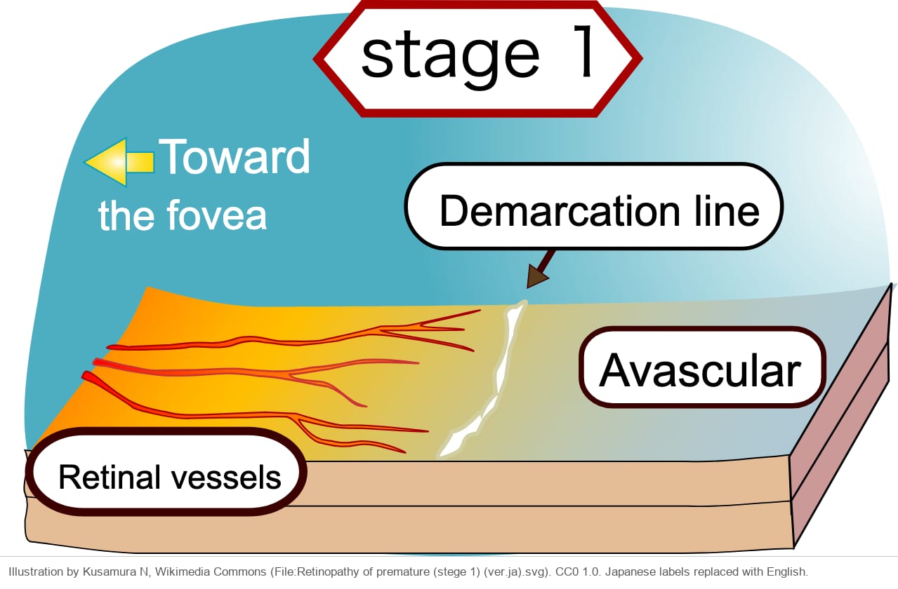
Stage 1: demarcation line
A thin, flat, white line between the vascularized and the avascular retina. It lies within the plane of the retina. The vessels just behind it may branch abnormally.
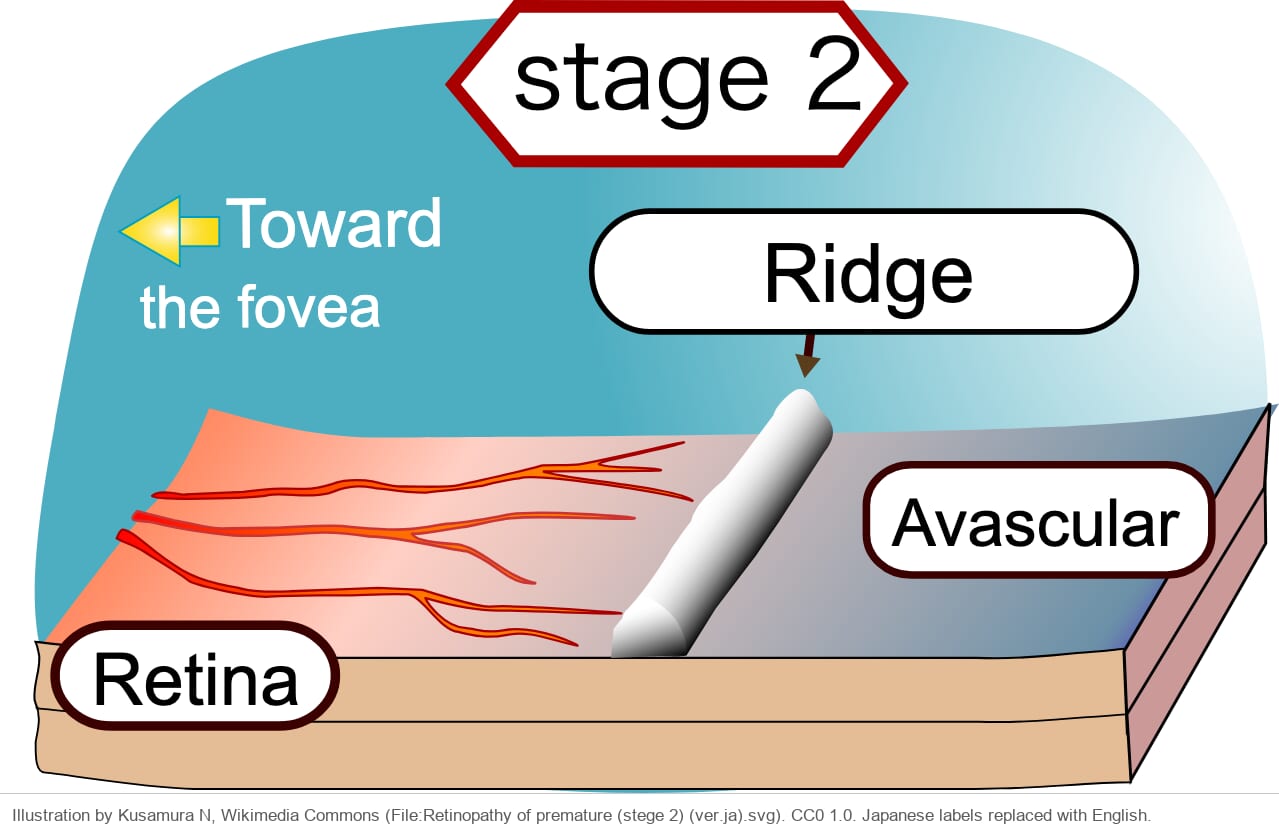
Stage 2: ridge
The line grows into a ridge with height and width. Small isolated tufts of new vessels, called "popcorn", may sit behind the ridge. Popcorn alone is still stage 2[2].
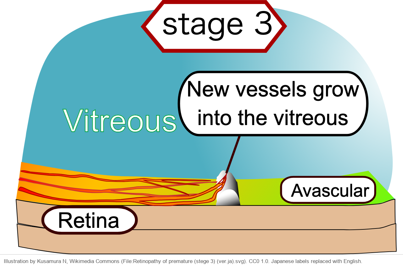
Stage 3: new vessels into the vitreous
New vessels grow from the ridge into the vitreous and give the ridge a ragged look. In zone I or posterior zone II, the new vessels can lie flat on the retina without a clear ridge. This is still stage 3.
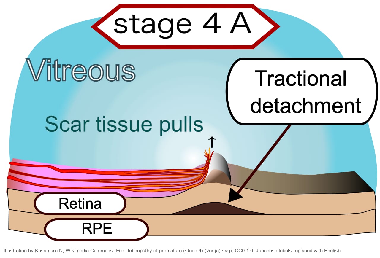
Stage 4: partial retinal detachment
The fibrovascular tissue contracts and pulls the retina off. Stage 4A spares the fovea, and stage 4B involves it.
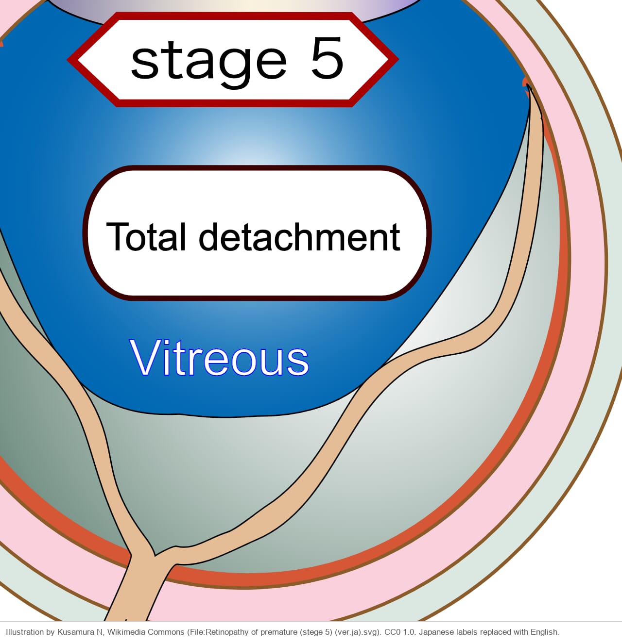
Stage 5: total retinal detachment
The whole retina is detached. ICROP3 divides it into 5A (the disc is visible), 5B (the disc is hidden behind retrolental fibrovascular tissue or a closed funnel) and 5C (5B with anterior segment changes such as a very shallow anterior chamber)[2].
Plus disease
As ROP gets worse, the posterior veins dilate and the arteries become tortuous. This is plus disease, a sign of active disease that is likely to progress. Vessels that are abnormal but not yet enough for plus are called pre-plus.
Aggressive ROP (A-ROP)
A-ROP is a severe, rapidly progressing form of ROP. ICROP3 renamed aggressive posterior ROP (AP-ROP) to A-ROP, because it also occurs in larger babies and outside the posterior retina, especially where resources are limited.
Usual ROP starts in the periphery and moves through stage 1, 2 and 3. A-ROP can skip this sequence. Severe plus disease and new vessels appear quickly, often with shunt vessels between arteries and veins that look like dilated loops. The new vessels can be flat and easy to miss with a 28-diopter lens. A lens with more magnification, such as a 20-diopter lens, or fluorescein angiography helps[2].
Untreated, A-ROP usually reaches stage 5, sometimes within a few days. Do not wait for the usual stages. Treat it promptly.
When to treat
The treatment criteria used worldwide come from the Early Treatment for ROP (ETROP) trial. They use zone, stage and plus disease to define two groups: one to treat and one to watch closely.
Type 1 ROP is any one of these three. Treat it within 72 hours.
- Zone I, any stage, with plus disease
- Zone I, stage 3, without plus disease
- Zone II, stage 2 or 3, with plus disease
Type 2 ROP is either of these two. Watch it closely.
- Zone I, stage 1 or 2, without plus disease
- Zone II, stage 3, without plus disease
About 10% of screened babies need treatment[1].
Laser or anti-VEGF
Laser photocoagulation treats the avascular peripheral retina, which makes the VEGF. Intravitreal anti-VEGF drugs such as bevacizumab and ranibizumab are also used. As a general rule, anti-VEGF is the first choice for zone I ROP and laser for zone II ROP. With anti-VEGF, the vessels can keep growing into the periphery, and the eye is less likely to become highly myopic. But ROP can come back after the drug wears off[1].
Stages 4 and 5 need surgery. Even when the retina is reattached, vision in stage 4B and 5 is usually poor[1].
After the acute phase
Regression
The first sign of regression is in the vessels: plus disease settles. It starts within 1–3 days after anti-VEGF and about 7–14 days after laser. The new vessels on the ridge become thin and white[2].
Long-term problems
Scarring is worse when the disease was more advanced and more posterior[1]. ICROP3 lists the long-term problems seen after ROP[2]:
- Vascular changes: straightened vessels with the macula and disc dragged temporally, or a falciform retinal fold
- Avascular retina that never vascularized (persistent avascular retina), which can thin and form holes
- Retinal detachment years later
- Angle-closure glaucoma
Children with ROP also need follow-up for refractive error, strabismus and amblyopia. Myopia is common, and high myopia is more likely after laser than after anti-VEGF[1].
Summary
- Describe every eye with ROP by zone, stage and plus disease (ICROP3).
- Zone I is the most posterior and the most dangerous zone.
- Stage 1 is a line, stage 2 a ridge, stage 3 new vessels into the vitreous, stage 4 a partial and stage 5 a total detachment.
- Judge plus disease from the zone I vessels, not by counting quadrants.
- Treat type 1 ROP within 72 hours: zone I with plus, zone I stage 3, or zone II stage 2 or 3 with plus.
- A-ROP skips the usual stages. After anti-VEGF, watch for reactivation.
Figure credits
Zones: Kusamura N, Wikimedia Commons, File:Zone1.2.3.svg. Public domain. Unchanged except for cropping the margins.
Stages: Kusamura N, Wikimedia Commons, File:Retinopathy of premature (stege 1, 2, 3, 4 and 5) (ver.ja).svg. CC0 1.0. Japanese labels replaced with English.
References
[1] Salmon JF. Kanski’s Clinical Ophthalmology: A Systematic Approach. 10th ed. Elsevier; 2025. Chapter 13, Retinal vascular disease: Retinopathy of prematurity.
[2] Chiang MF, Quinn GE, Fielder AR, et al. International Classification of Retinopathy of Prematurity, Third Edition. Ophthalmology. 2021;128(10):e51-e68. PMID 34247850.