Uveitis at the First Visit: How to Narrow Down the Cause
Uveitis has a reputation for being hard. It has many causes, and even after a full work-up in specialist centers many patients never get a named diagnosis. In published series, 38 to 56% of anterior uveitis is idiopathic, and 36.6% of 5,378 patients in a Japanese nationwide survey were left unclassified[1]. Still, start every case by looking for the cause. Treating every anterior chamber reaction with steroid drops without asking why does not make you better at uveitis, and it can make an infection worse.
Thinking carefully about the findings and the epidemiology narrows down the causes to some extent. This article goes through five questions, then who needs tests and what not to miss.
Where is the inflammation?
You usually suspect uveitis when you see cells in the anterior chamber. Grade them before you dilate, because dilating can release pigment into the aqueous. Then look at the anterior vitreous and the fundus, and place the inflammation:
- Anterior uveitis: the anterior chamber is the main site of inflammation.
- Posterior uveitis: the inflammation is mainly in the retina or choroid.
- Panuveitis: no single main site; the anterior chamber, the vitreous and the retina or choroid are all inflamed.
There is also intermediate uveitis, where the vitreous is the main site. It is less common, and this article leaves it aside.
Sometimes the only finding is cells in the anterior vitreous, with no cells in the anterior chamber and no chorioretinal lesion. In that case, before thinking of uveitis, look for pigment from a retinal tear (tobacco dust) or a vitreous hemorrhage, and examine the peripheral retina.
Acute, recurrent or chronic?
Next, ask how the inflammation began and how long it has lasted. Acute anterior uveitis starts suddenly and settles within 3 months. It gives a painful red eye with photophobia and blurred vision, ciliary injection and a small pupil. When acute attacks come back with 3 months or more of a quiet eye off treatment in between, the course is called recurrent.
Chronic anterior uveitis lasts longer, or comes back within 3 months of stopping treatment. It is more often bilateral and more often has a systemic cause. Granulomatous signs are common, and the eye can be white with no symptoms until complications such as cataract appear.
One eye or both?
Bilateral uveitis is more often immune-mediated (sarcoidosis, Vogt-Koyanagi-Harada disease), and unilateral uveitis more often infectious (endophthalmitis, herpes). This is a tendency, with exceptions.
Two cautions. A bilateral disease can show signs in only one eye at the first visit. And mild inflammation in the fellow eye is easy to miss, so examine the "normal" eye as carefully as the red one. HLA-B27 acute anterior uveitis is the classic exception: it is immune-mediated, but it affects one eye at a time. Either eye can be affected at different times, and both together is rare.
Granulomatous or non-granulomatous?
The difference is in the cells. In granulomatous inflammation, macrophages and lymphocytes lead, and they tend to clump together. In non-granulomatous inflammation, neutrophils lead, and they stay scattered. Sarcoidosis is the typical granulomatous disease, Behçet disease the typical non-granulomatous one. Each sign follows from this.
Keratic precipitates
In granulomatous inflammation the cells clump into large, greasy keratic precipitates, called mutton-fat KP. They settle on the lower cornea under gravity. In non-granulomatous inflammation the KP are fine.
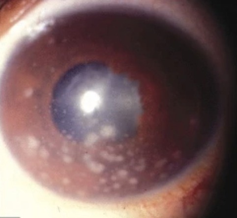
Large white KP are scattered over the cornea, and they are most crowded on the lower half.
Hypopyon
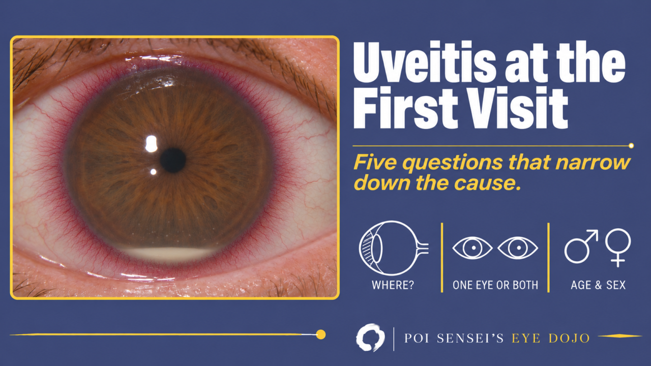
In Behçet disease the hypopyon is made of neutrophils with little fibrin, so it is fluid and shifts when the patient tilts the head. In HLA-B27 acute anterior uveitis, endophthalmitis and infectious keratitis, the hypopyon contains fibrin, so it does not move and can even heap up.
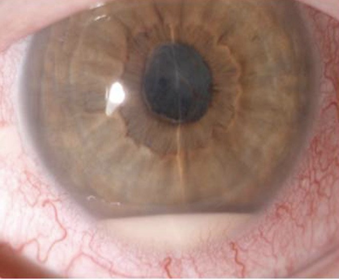
The hypopyon sits at the bottom of the anterior chamber with a smooth, level top.
Fundus
In sarcoidosis the cells clump in the vitreous into snowballs. Clumps are heavy and sink, so snowballs gather in the lower vitreous. When you suspect sarcoidosis, always look at the inferior retina.
Do not rely on this split alone
Not every disease fits. Syphilis can look granulomatous or non-granulomatous. Bacterial endophthalmitis shows fibrin and hypopyon, which fit non-granulomatous inflammation, together with mutton-fat KP. Use the split to put the possibilities in order.
Age and sex
Age and sex narrow the list further. This table lists the diseases that can show anterior chamber inflammation at the first visit.
| Disease | Typical age | Sex |
|---|---|---|
| HLA-B27 acute anterior uveitis | Young adults | Male |
| Behçet disease | 20s–40s | Eye disease: male |
| Sarcoidosis | Young and middle-aged adults | Varies between studies |
| Vogt-Koyanagi-Harada disease | 10s–40s | Female |
| Posner-Schlossman syndrome | 20s–50s | Slightly male |
| Herpetic anterior uveitis | Older | None |
| Diabetic iritis | Any | None |
| Syphilitic uveitis | 20s–50s | Mostly male |
| Bacterial endophthalmitis | Older | Endogenous: slightly male |
| Intraocular lymphoma | Over 60 | Slightly female |
| TINU syndrome | 10s–30s | Female |
| Juvenile idiopathic arthritis | 1–12 years | Female |
How common each disease is depends on where you work and on the patient’s ancestry. HLA-B27 is carried by 6 to 8% of White people in the USA but by about 0.5% of Japanese people. Vogt-Koyanagi-Harada disease is common in Japan and Latin America and in people of Asian, Hispanic or Native American descent, but very rare in people of northern European descent. In the USA, sarcoidosis is more than three times as common in African Americans as in European Americans. Behçet disease is most common along the old Silk Road, from Turkey through the Middle East to the Far East.
A few diseases in the table have a sign that helps at the first visit:
- Herpetic anterior uveitis: raised intraocular pressure (IOP) and patchy or sectoral iris atrophy. Recurrences almost always involve the same eye. Ask about previous herpes simplex keratitis, cold sores or shingles around the eye.
- Posner-Schlossman syndrome: IOP often above 40 mmHg with only a few cells and fine KP, and no posterior synechiae.
- Diabetic iritis: very high blood glucose.
- TINU syndrome (tubulointerstitial nephritis and uveitis): adolescent girls, usually after the kidney disease.
Putting it together
No single answer gives the diagnosis, but together they often point to one disease. Here is the most common example.
A young man notices pain and blurred vision in one eye. There is marked ciliary injection, many anterior chamber cells and fine KP, often with fibrin or a hypopyon that does not move. The answers are anterior, acute, one eye, non-granulomatous, young and male. This is the classic HLA-B27 acute anterior uveitis.
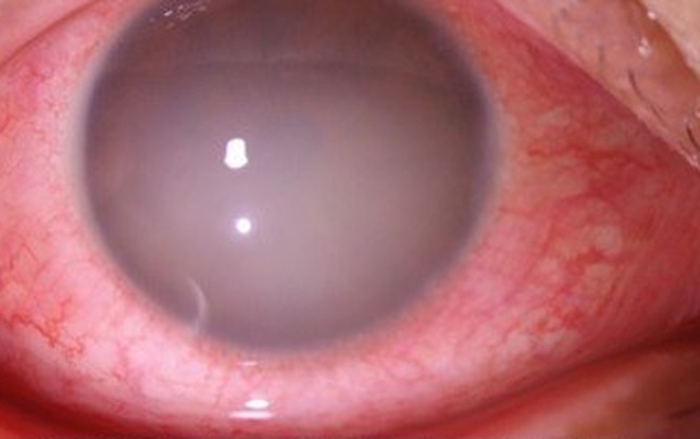
The redness is strongest around the cornea, and the anterior chamber is so cloudy with protein that the iris is hard to see.
So ask about low back or buttock pain and morning stiffness, which point to ankylosing spondylitis. The back pain can start after the eye attack. If a patient you are following mentions back pain, think of it again.
Two other diseases can also give a unilateral, non-granulomatous inflammation with fibrin and a hypopyon: diabetic iritis and bacterial endophthalmitis. So even when you think it is acute anterior uveitis, always think about a fundus examination and a blood glucose test. Once you know the epidemiology and the typical case, learn the diseases that look like it. This is how you get better at differential diagnosis.
Who needs tests?
Looking for the cause is not the same as ordering tests. A first episode of acute anterior uveitis usually needs no tests for a systemic cause when all of these apply:
- one eye
- mild to moderate
- non-granulomatous
- no hypopyon
- nothing in the eye or the history that suggests a cause
The fundus examination and blood glucose test above are separate from this: think about them in every patient.
Order tests when the uveitis is:
- recurrent, severe or bilateral
- chronic or not settling with treatment
- granulomatous
- accompanied by intermediate or posterior uveitis
- accompanied by any eye or systemic clue to a cause
Choose the tests from the findings rather than ordering a fixed panel. HLA-B27 is worth testing in recurrent or chronic non-granulomatous anterior uveitis. For syphilis, order both a treponemal test and a non-treponemal test (RPR or VDRL). For sarcoidosis, add serum ACE and lysozyme and a chest X-ray. For tuberculosis, add an interferon-gamma release assay.
Do not miss
Some diseases cause permanent loss of vision if treatment is late. Bacterial endophthalmitis leads the list after intraocular surgery, and endogenous endophthalmitis can look like a fibrinous anterior uveitis. Acute retinal necrosis comes with marked anterior chamber and vitreous inflammation, so look for patches of necrosis in the peripheral retina. Syphilis can look like almost any uveitis.
If you have settled on a less urgent diagnosis but something does not fit, see the patient again the next day or within a few days, not in a week.
Summary
- Place the inflammation first. Cells only in the anterior vitreous: think of a retinal tear or hemorrhage before uveitis.
- Bilateral uveitis is more often immune-mediated and unilateral uveitis more often infectious, but examine both eyes.
- Mutton-fat KP suggest granulomatous inflammation, and a mobile hypopyon suggests Behçet disease.
- A young man with unilateral fibrinous anterior uveitis is the typical HLA-B27 patient. Ask about back pain.
- If something does not fit your diagnosis, see the patient again within a few days.
References
[1] Sonoda KH, Hasegawa E, Namba K, et al. Epidemiology of uveitis in Japan: a 2016 retrospective nationwide survey. Jpn J Ophthalmol. 2021;65(2):184-190. PMID 33694024.
Which retinal layer makes which wave, the three abnormal patterns, and the dark-adaptation trap to check before you report “negative” — on one page, by email. One email when I publish something new, about once a month. Unsubscribe anytime.