Familial Exudative Vitreoretinopathy (FEVR): Fundus Signs, Stages and Pseudostrabismus
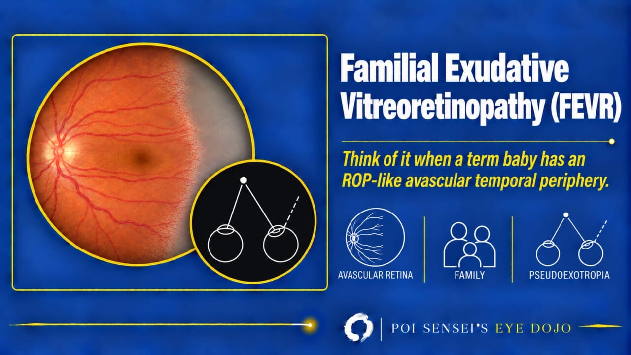
A baby born at term can have a retina that looks like retinopathy of prematurity (ROP). That is the clue to familial exudative vitreoretinopathy (FEVR). This article shows the fundus and angiography signs, the stages, and why a child with FEVR can look exotropic without a true squint.
- A child born at term has an avascular temporal periphery, like ROP.
- A child or young adult has a retinal detachment, especially with a family history.
- A child has leukocoria.
- A child looks exotropic, but the cover test shows no movement.
What is FEVR?
In FEVR, the retinal vessels fail to grow all the way to the temporal periphery, as in ROP. The avascular retina is hypoxic and drives new vessels. These can lead to vitreoretinal traction, exudation, hemorrhage and tractional retinal detachment[1].
Inheritance is most often autosomal dominant, and rarely X-linked or autosomal recessive. The two eyes are often affected unequally, and severity can vary a lot even within one family. Only 20–40% of patients have a known family history, so a negative history does not rule FEVR out[3].
Fundus and angiography
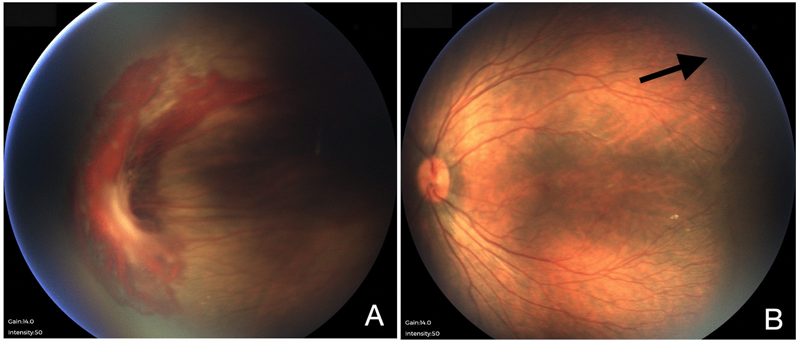
Look at the temporal periphery. The typical signs are:
- Retinal vessels that stop abruptly at the temporal equator, with avascular retina beyond
- Straightened vessels running toward the temporal periphery
- Many small branches and dilated small vessels (telangiectasia) at the vascular edge
- Dragging of the disc and macula toward the temporal side
- In more advanced eyes, exudates, sickle-shaped retinal folds (falciform folds) and retinal detachment
High myopia may be present[1][2].
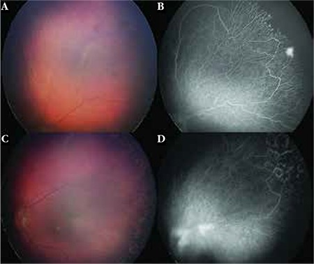
Wide-field fluorescein angiography (FA) is the key test. Mild FEVR can look nearly normal on fundus examination. FA shows the avascular periphery, the abnormal branching and loops at the vascular edge, and leakage[1][3]. It also shows which avascular retina to treat with laser.
Stages
The revised classification (2014) has five stages. Stages 1 to 4 are split into A, without exudate or leakage on FA, and B, with exudate or leakage[3][4].
| Stage | Finding |
|---|---|
| 1 | Avascular periphery or abnormal vessels within the retina |
| 2 | New vessels growing from the retina into the vitreous |
| 3 | Retinal detachment not involving the macula |
| 4 | Retinal detachment involving the macula |
| 5 | Total retinal detachment |
FEVR does not always pass through the stages in order[3].
FEVR or ROP?
The fundus can look the same. The history helps. Children with FEVR were born at term, or their disease does not follow the time course of ROP[3]. FEVR can also progress after years of quiet[1].
When to suspect FEVR
Leukocoria
An eye with a total retinal detachment shows a white pupil. Keep FEVR in the differential with ROP, Coats disease, Norrie disease and retinoblastoma.
Retinal detachment in a child or young adult
Most detachments in FEVR occur before age 10. They are caused by traction in early childhood, and by both traction and retinal breaks in the teens[2]. In a young patient with a detachment, look at the periphery of the other eye, and ask about the family.
Family members
Relatives without symptoms often have stage 1 or 2 disease[3]. Examine them, ideally with wide-field angiography.
FEVR and strabismus
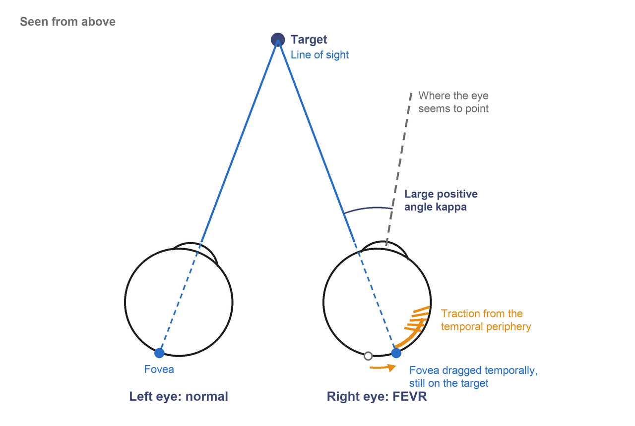
Traction can pull the macula toward the temporal side. An ectopic macula is found in about half of patients[2].
The patient still fixates with the fovea. Because the fovea has moved temporally, the eye must turn out to point it at a target. The corneal light reflex then sits nasally (a large positive angle kappa), and the eye looks exotropic[1][2].
Treatment and follow-up
Stage 1 without leakage needs no treatment, only regular follow-up[3]. When there are new vessels or leakage on FA, especially at the junction of the vascularized and avascular retina, laser the avascular retina[1][4][5]. Retinal detachment needs surgery, such as vitrectomy or scleral buckling. The vitreous sticks firmly to the avascular retina, so the surgery is difficult[1][5].
FEVR tends to progress in childhood and settle after the second decade. But it can worsen at any age after years of quiet, so follow-up is lifelong[1].
Summary
- FEVR looks like ROP in a baby born at term: avascular temporal periphery, abrupt vessel ends and straightened vessels.
- It is usually autosomal dominant, but a negative family history does not rule it out.
- Wide-field FA shows the avascular periphery and leakage, and where to laser.
- Stages 1 to 5: avascular retina, new vessels, detachment without and with the macula, total detachment.
- A dragged macula makes the eye look exotropic, but the cover test shows no movement.
- Laser the avascular retina when there are new vessels or leakage, examine the family, and follow for life.
Figure credits
Fundus photographs: Fig. 3 from Wai YZ, Chong YY, Lim LT, et al. Int J Retina Vitreous 2022;8:30, licensed under CC BY 4.0. Unchanged.
Fluorescein angiography: Figure 2 from Sızmaz S, Yonekawa Y, Trese MT. Turk J Ophthalmol 2015;45:164-168, licensed under CC BY 2.5. Unchanged.
The pseudoexotropia drawing is my own.
References
[1] Salmon JF. Kanski’s Clinical Ophthalmology: A Systematic Approach. 10th ed. Elsevier; 2025. Chapter 15, Hereditary fundus dystrophies: Familial exudative vitreoretinopathy; Chapter 18, Strabismus: Pseudostrabismus.
[2] Yanoff M, Duker JS. Ophthalmology. 5th ed. Elsevier; 2019. Chapter 6.17, Hereditary vitreoretinopathies.
[3] Sızmaz S, Yonekawa Y, Trese MT. Familial exudative vitreoretinopathy. Turk J Ophthalmol. 2015;45(4):164-168. PMID 27800225.
[4] Kashani AH, Brown KT, Chang E, Drenser KA, Capone A, Trese MT. Diversity of retinal vascular anomalies in patients with familial exudative vitreoretinopathy. Ophthalmology. 2014;121(11):2220-2227. PMID 25005911.
[5] Meier P, Wiedemann P. Surgery for pediatric vitreoretinal disorders. In: Ryan’s Retina. Elsevier. Chapter 118.