Geographic Atrophy on OCT: cRORA, iRORA and Nascent GA, and What Treatment Changes
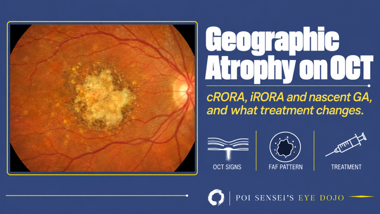
Geographic atrophy (GA) is the late stage of dry age-related macular degeneration (AMD). It is common in White patients and less common in East Asia, including Japan. Two complement inhibitors have now reached clinics. This article shows how to call GA on optical coherence tomography (OCT), how to tell it from other macular atrophy, what fundus autofluorescence (FAF) adds, and what the new injections can and cannot do.
- On OCT, does the atrophy meet the four signs of complete RPE and outer retinal atrophy (cRORA)?
- Is there any sign of present or past macular neovascularization (MNV)?
- On FAF, how large is it, and is it one lesion or many? On near-infrared or OCT, does it reach the fovea?
- If you treat, is the lesion away from the fovea?
What GA is
Drusen build up under the retinal pigment epithelium (RPE). Over years, the RPE dies, along with the photoreceptors above it and the choriocapillaris below it. The dead area spreads slowly. Most GA appears where large drusen used to be. When it reaches the fovea, central vision is lost for good. Even before that, a ring of atrophy around a spared fovea makes reading hard.
Complement, part of the immune system, drives much of this damage. That is why the new drugs block complement proteins C3 or C5.
Calling GA on OCT
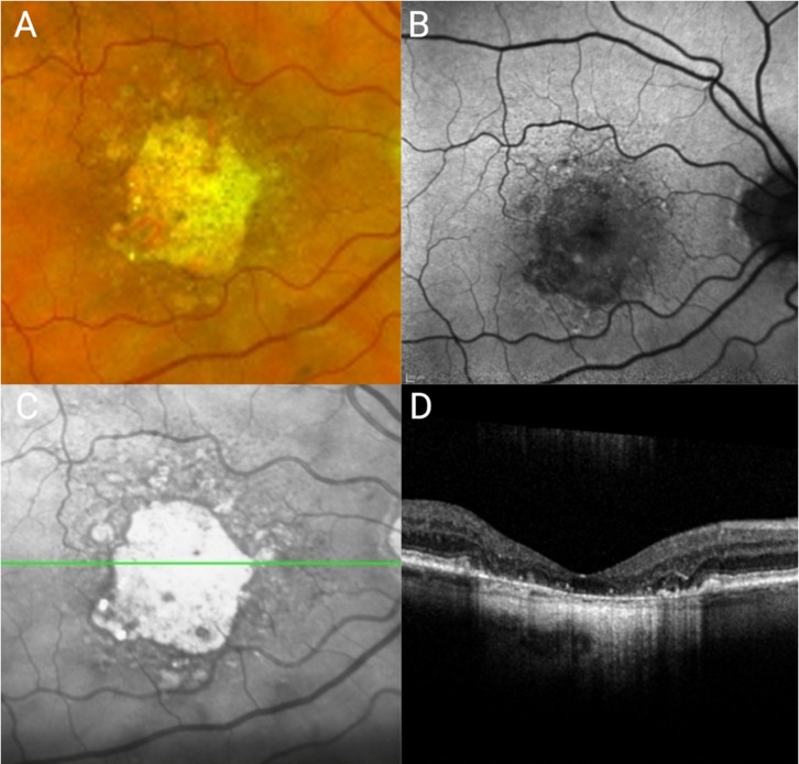
Color photos and FAF show GA well, but OCT now sets the definition. The Classification of Atrophy Meetings (CAM) gave OCT names to each stage of atrophy. In short, cRORA (complete RPE and outer retinal atrophy) is the OCT name for complete atrophy, and GA is cRORA in an eye with no present or past MNV.
cRORA: complete RPE and outer retinal atrophy
All four must be present on a B-scan:
- Hypertransmission at least 250 µm wide. Light passes through the missing RPE and lights up the choroid below.
- RPE loss or thinning at least 250 µm wide.
- Photoreceptor loss above it: the ellipsoid zone, interdigitation zone and external limiting membrane are gone, and the outer nuclear layer is thin.
- No RPE tear. A scrolled RPE edge means a tear, not atrophy.
The 250 µm cut-off is a consensus number, not a biological line, and it may change[1]. Do not judge from the foveal scan alone. Take a volume scan (many B-scans across the macula) and measure on the B-scan that runs through the widest part of the lesion.
iRORA: incomplete RPE and outer retinal atrophy
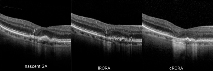
iRORA has the first three signs, but not enough to meet cRORA: for example, the hypertransmission or RPE loss is under 250 µm. Hypertransmission is limited, the RPE is partly preserved, and the photoreceptors are starting to fail. It is atrophy that has started but is not yet complete. Most iRORA turns into cRORA: in one series, 93.1% of lesions did so within 24 months, with a median of 14 months[3].
Why split the two? cRORA is expected to give a dense scotoma, while iRORA still keeps some sensitivity. The split also gives trials an earlier target: if treatment can slow the step from iRORA to cRORA, it may save more retina.
Nascent GA
Before the RPE breaks down, OCT can show the outer retina sinking. The outer plexiform layer and inner nuclear layer subside, and a dark wedge appears in the Henle fiber layer. This was first named nascent GA (nGA), and atrophy followed on average 11 months (range 5 to 21) later[4]. CAM later widened the term: nGA now means iRORA in an eye with no present or past MNV[2]. So today nGA and iRORA largely overlap, although some figures, like the one below, still draw nGA as the earliest step. You will not hear the term often in clinics.
When cRORA is called GA
GA is one kind of cRORA. Call it GA only when the atrophy is not explained by something else:
- present or past MNV
- a laser scar
- inflammation
- an inherited retinal disease such as Stargardt disease or central areolar choroidal dystrophy
Atrophy that is not GA
Three kinds of atrophy can look like GA.
Macular atrophy after neovascular AMD
Eyes treated for neovascular AMD often develop atrophy over years of injections. It can meet every cRORA sign. But the cause is different: the MNV regresses or scars, and anti-VEGF treatment may add to it. When there is present or past MNV, CAM asks us to say cRORA on OCT and macular atrophy on the fundus, not GA.
Pachychoroid GA
In Japan, some GA-like atrophy sits on a thick choroid with no drusen. In a Japanese study, these patients were younger and mostly men, and usually had a single small lesion that grew more slowly than conventional GA[5]. The atrophy likely comes from poor choroidal flow, not from drusen. There are no data yet on whether complement inhibitors help these eyes.
EMAP
Extensive macular atrophy with pseudodrusen-like appearance (EMAP) is a different disease. It starts in the 40s or 50s, often with night blindness. The atrophy is taller than it is wide, reaches the fovea early and grows fast, with pseudodrusen-like deposits across the fundus and paving-stone changes in the far periphery[6]. Think of it when a "GA" patient is young and the atrophy grows fast.
Speed is a clue in general. GA grows over months to years. Atrophy that spreads from week to week is not GA, so look for inflammation or another cause.
FAF: size and pattern
On FAF, GA is a dark area with a sharp border. Trials measure its area on FAF. Blue-light FAF has a blind spot at the fovea: macular pigment makes it look dark. Use near-infrared reflectance or OCT to decide whether the fovea is involved.
Patterns around the lesion
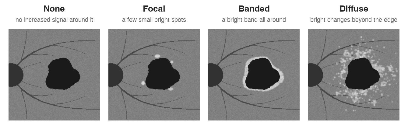
Just outside the lesion, FAF is often brighter than normal. This bright signal comes from abnormal RPE at the border of the atrophy. Its shape is sorted into patterns, and the figure shows the four common ones. In the FAM study, median growth was about 1.8 mm² a year with a banded or diffuse pattern, 0.8 mm² with a focal pattern and 0.4 mm² with none[7]. But bigger lesions add more mm² each year, so part of this gap is just lesion size. A 2026 study that adjusted for lesion size found that only the diffuse pattern grew a little faster, and about 1 in 5 eyes changed pattern within a few years[8]. Treat the pattern as a rough hint, not a forecast.
How fast it grows, and how to measure it
GA grows by about 1.5 mm² a year on average, but the range is wide. Large lesions and multifocal lesions grow faster in area, and lesions grow more slowly toward the fovea than away from it[9].
Raw area is a poor yardstick because of this size effect. Trials take the square root of the area, which turns it into a length and removes much of this effect. Growth is then given in mm/year.
When you follow a patient, compare like with like: the same device and the square-root area.
Treatment: complement inhibitors
Two intravitreal drugs are in use. Both slow lesion growth compared with sham (a simulated injection). Neither has improved vision in trials.
| Pegcetacoplan | Avacincaptad pegol | |
|---|---|---|
| Target | C3 | C5 |
| Dosing in trials | Monthly or every other month | Monthly |
| Slower growth vs sham | 16‑22% at 24 months | 14% at 12 months |
| New MNV vs sham | 6‑13% vs 2‑4% at 24 months | 7% vs 4% at 12 months |
For pegcetacoplan, OAKS showed the effect at 12 months, while DERBY reached significance only at 24 months. Visual function did not differ from sham at 24 months. GATHER2 tested avacincaptad pegol only in eyes with GA that did not involve the foveal center[11]. In both programs, new MNV was the main extra risk.
The US FDA approved both drugs in 2023. In Europe, the European Medicines Agency refused pegcetacoplan in 2024, because slower growth did not help patients in daily life. Japan gave avacincaptad pegol conditional approval in 2025.
Who to treat
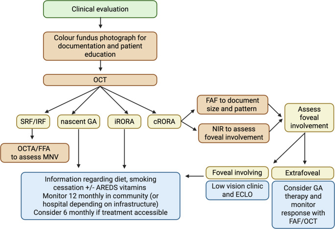
The usual candidate is cRORA without MNV that has not yet reached the foveal center. Here, slowing growth may delay atrophy from reaching the fovea. In OAKS and DERBY, the effect at 12 months was also larger in lesions away from the fovea (23-26%) than in lesions under it (8-11%)[10]. A later analysis of GATHER1 suggested that treatment may also slow iRORA turning into cRORA, but the numbers were small, and treating iRORA is still research[12].
Whatever you decide about injections, give the basics: stop smoking, antioxidant supplements when indicated, an Amsler grid, and low vision help when reading fails.
Summary
- Call GA on OCT with the four cRORA signs, and only after ruling out MNV and other causes.
- iRORA (called nascent GA when there is no MNV) comes before it and often progresses to cRORA.
- Atrophy after neovascular AMD, pachychoroid atrophy and EMAP are not GA.
- On FAF, check the size and whether it is one lesion or many. Check the fovea on near-infrared reflectance or OCT.
- Complement inhibitors slow growth by 14-22%, more away from the fovea, but add new MNV without better vision.
References
[1] Sadda SR, Guymer R, Holz FG, et al. Consensus definition for atrophy associated with age-related macular degeneration on OCT: Classification of Atrophy Report 3. Ophthalmology. 2018;125(4):537-548. PMID 29103793.
[2] Guymer RH, Rosenfeld PJ, Curcio CA, et al. Incomplete retinal pigment epithelial and outer retinal atrophy in age-related macular degeneration: Classification of Atrophy Meeting Report 4. Ophthalmology. 2020;127(3):394-409. PMID 31708275.
[3] Corradetti G, Corvi F, Nittala MG, et al. Natural history of incomplete retinal pigment epithelial and outer retinal atrophy in age-related macular degeneration. Can J Ophthalmol. 2021;56(5):325-334. PMID 33539821.
[4] Wu Z, Luu CD, Ayton LN, et al. Optical coherence tomography-defined changes preceding the development of drusen-associated atrophy in age-related macular degeneration. Ophthalmology. 2014;121(12):2415-2422. PMID 25109931.
[5] Sato Y, Ueda-Arakawa N, Takahashi A, et al. Clinical characteristics and progression of pachychoroid and conventional geographic atrophy. Ophthalmol Sci. 2024;4(5):100528. PMID 38827489.
[6] Hamel CP, Meunier I, Arndt C, et al. Extensive macular atrophy with pseudodrusen-like appearance: a new clinical entity. Am J Ophthalmol. 2009;147(4):609-620. PMID 19181301.
[7] Holz FG, Bindewald-Wittich A, Fleckenstein M, et al. Progression of geographic atrophy and impact of fundus autofluorescence patterns in age-related macular degeneration. Am J Ophthalmol. 2007;143(3):463-472. PMID 17239336.
[8] Antropoli A, Vacirca F, Introini U, et al. Perilesional fundus autofluorescence patterns are not static: longitudinal transitions in geographic atrophy and association with disease progression. Ophthalmol Sci. 2026;6(2):100995. PMID 41439216.
[9] Borchert GA, Charbel Issa P, Xue K, et al. Geographic atrophy in age-related macular degeneration: phenotypic characterisation for clinical trial consideration. Graefes Arch Clin Exp Ophthalmol. 2026;264(5):1201-1216. PMID 41721855.
[10] Heier JS, Lad EM, Holz FG, et al. Pegcetacoplan for the treatment of geographic atrophy secondary to age-related macular degeneration (OAKS and DERBY): two multicentre, randomised, double-masked, sham-controlled, phase 3 trials. Lancet. 2023;402(10411):1434-1448. PMID 37865470.
[11] Khanani AM, Patel SS, Staurenghi G, et al. Efficacy and safety of avacincaptad pegol in patients with geographic atrophy (GATHER2): 12-month results from a randomised, double-masked, phase 3 trial. Lancet. 2023;402(10411):1449-1458. PMID 37696275.
[12] Corradetti G, Karamat A, Srinivas S, et al. Progression to complete retinal pigment epithelium and outer retinal atrophy (cRORA): post hoc analysis of the GATHER1 trial. Graefes Arch Clin Exp Ophthalmol. 2025;263(3):669-677. PMID 39538001.
Which retinal layer makes which wave, the three abnormal patterns, and the dark-adaptation trap to check before you report “negative” — on one page, by email. One email when I publish something new, about once a month. Unsubscribe anytime.