Central Areolar Choroidal Dystrophy (CACD): Fundus, Autofluorescence and ERG
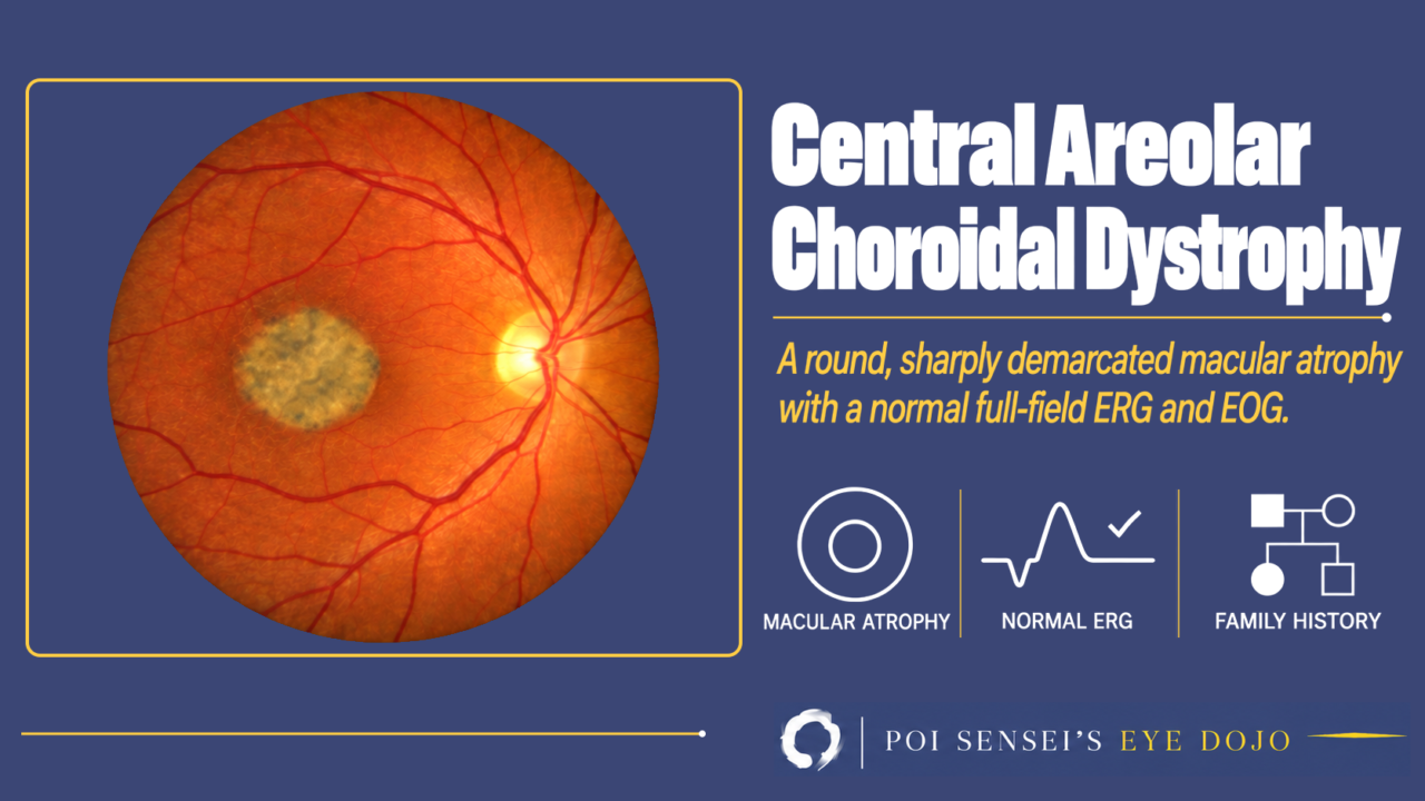
Both eyes have a round, sharply demarcated patch of atrophy at the macula, and the patient is an adult with slowly falling central vision. Think of central areolar choroidal dystrophy (CACD). This article shows what CACD looks like on the fundus, autofluorescence and OCT, why the electroretinogram (ERG) still matters, and how not to mistake it for late Best disease or dry age-related macular degeneration (AMD).
- Central vision slowly falls in adult life.
- Both maculas show a symmetric, round, sharply demarcated patch of atrophy.
- Fundus autofluorescence shows a dark patch, often with a bright ring around it.
- The full-field ERG is usually normal.
What is CACD?
CACD is an inherited macular dystrophy. The retinal pigment epithelium (RPE) and the choriocapillaris at the macula waste away, and a round area of atrophy forms in both eyes.
Symptoms and inheritance
Central vision slowly gets worse, usually starting between the ages of 30 and 60. This is later than in most other macular dystrophies[1]. If the fovea is still spared, visual acuity can stay good for a while.
Most families show autosomal dominant inheritance. The gene most often involved is PRPH2.
Findings
Fundus
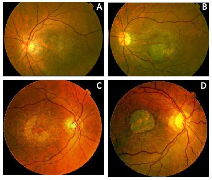
Both maculas show a symmetric, sharply demarcated, round patch of atrophy. The disease goes through four stages[1]:
- Stage I: slight pigment changes around the fovea.
- Stage II: a round, pale, mildly atrophic area at the macula.
- Stage III: well-defined atrophy next to the fovea, with the center spared.
- Stage IV: the atrophy covers the fovea, and the large choroidal vessels show through it.
It often takes more than ten years to move from stage II to stage III. Vision falls sharply at stage IV.
Fundus autofluorescence
Fundus autofluorescence is very useful for the diagnosis. Early lesions show a speckled mix of bright and dark spots. Once the RPE is lost, the atrophic area is dark (hypoautofluorescent). In advanced disease, a thin bright (hyperautofluorescent) ring often remains around it[1].
Fluorescein angiography
On fluorescein angiography, early lesions are bright from a window defect: the choroidal fluorescence shows through the thinned RPE. Once the choriocapillaris is lost, the area turns dark and the large choroidal vessels stand out. A little dye may leak at the edge of the atrophy. Unlike wet AMD, there is usually no leakage from new vessels, and choroidal neovascularization is rare.
OCT
OCT shows loss of the outer retina, including the photoreceptors, and the RPE in the atrophic area.
ERG
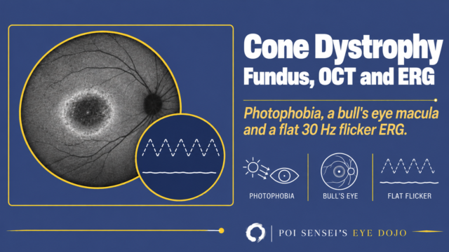
The full-field ERG records the response of the whole retina, so a disease limited to the macula hardly changes it. In CACD, the full-field ERG is usually normal, even at stage IV. A few patients have a reduced cone response. The multifocal ERG, which maps responses across the macula, is reduced there[1].
A normal full-field ERG helps separate CACD from cone and cone-rod dystrophies, which also cause macular atrophy.
EOG
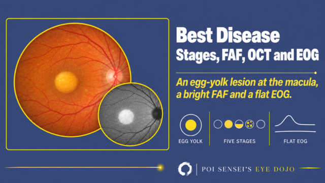
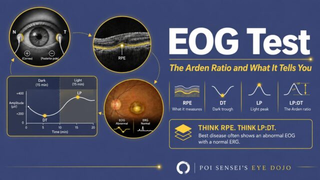
Late Best disease can look very similar to CACD: once the yellow material is gone, a round patch of atrophy is left at the macula. The electro-oculogram (EOG) helps tell them apart. In Best disease, the EOG is abnormal at every stage. In CACD, it is usually normal, though it can be reduced in advanced disease[1].
Treatment
There is no treatment that stops the disease. Low-vision care helps patients use their remaining vision.
Summary
- CACD is an inherited macular dystrophy, most often caused by PRPH2.
- Both maculas show symmetric, round, sharply demarcated atrophy, dark on autofluorescence.
- A normal full-field ERG helps separate it from cone and cone-rod dystrophies, and a normal EOG from late Best disease.
- In older patients it can be mistaken for geographic atrophy in AMD, so ask about the family.
References
[1] Boon CJ, Klevering BJ, Cremers FP, Zonneveld-Vrieling MN, Theelen T, Den Hollander AI, Hoyng CB. Central areolar choroidal dystrophy. Ophthalmology. 2009;116(4):771-782. PMID 19243827.
[2] Kersten E, Geerlings MJ, Pauper M, Corominas J, Bakker B, Altay L, Fauser S, de Jong EK, Hoyng CB, den Hollander AI. Genetic screening for macular dystrophies in patients clinically diagnosed with dry age-related macular degeneration. Clin Genet. 2018;94(6):569-574. PMID 30215852.
Which retinal layer makes which wave, the three abnormal patterns, and the dark-adaptation trap to check before you report “negative” — on one page, by email. One email when I publish something new, about once a month. Unsubscribe anytime.