Peripheral Exudative Hemorrhagic Chorioretinopathy (PEHCR): Findings, Imaging and How to Tell It From Melanoma
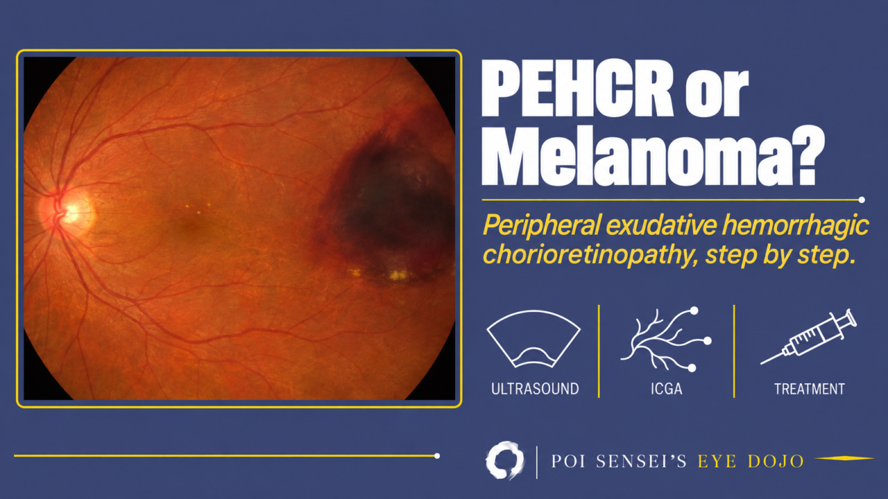
An older patient is referred with a dark, raised mass in the far temporal fundus. The referral letter says "choroidal melanoma." Before you plan treatment for a tumor, think of peripheral exudative hemorrhagic chorioretinopathy (PEHCR). This article explains what PEHCR is, how it looks on the fundus, ultrasound, OCT and angiography, how to tell it from melanoma, and when to treat it.
- An older adult has blood, lipid exudate or a pigment epithelial detachment (PED) in the peripheral fundus, usually on the temporal side.
- The dark "mass" is a hemorrhagic PED or a clot under the retina.
- Lipid exudate lies next to the blood.
- Drusen or other age-related changes are seen at the macula of either eye.
What is PEHCR?
Peripheral exudative hemorrhagic chorioretinopathy (PEHCR) is a degenerative disorder of the peripheral fundus in older adults. Blood, exudate or both collect under the retina and under the retinal pigment epithelium (RPE). Blood under the RPE lifts it off Bruch membrane as a dome, a pigment epithelial detachment (PED). It looks like the bleeding and exudation of neovascular age-related macular degeneration (AMD), but it sits outside the macula. Older names include eccentric disciform degeneration and peripheral disciform degeneration.
PEHCR is uncommon. Most eyes keep good vision, but vision is poor if the blood or fluid reaches the macula.
Who gets it
In the largest series of 173 eyes, the mean age was 80 years and two-thirds were women[1]. Most reports come from white patients, but PEHCR also occurs in Asian patients, sometimes at a younger age. A Korean series of 43 eyes had a mean age of 70[2].
Most patients have it in one eye. In the larger series, both eyes are involved in about a quarter to a third of patients[3]. The other eye can also hold a silent lesion, fresh or old and scarred, so look at its periphery too.
Half of the patients had high blood pressure, and 44% took aspirin or another blood thinner[1].
What causes it
The exact cause is unknown. In the broad sense, though, I think of PEHCR as neovascular AMD in the periphery. New vessels grow from the choroid under the RPE, and they bleed and leak. In many eyes the new vessels take the form of polypoidal choroidal vasculopathy (PCV), in which the vessels end in small balloon-like swellings (polypoidal lesions). Indocyanine green angiography (ICGA) shows this picture in the periphery of many eyes with PEHCR, and in some eyes peripheral PCV is clearly the source of the bleeding[4].
Where do the new vessels come from? There are two ideas, and both may be true in different eyes.
One is drusen-driven, as in typical AMD. The age is the same as AMD, and about half of the eyes have drusen, RPE changes or neovascularization at the macula.
The other is pachychoroid-driven, as in PCV and central serous chorioretinopathy: a thick, congested choroid drives the disease. In eyes with PEHCR, the choroid is thicker in the temporal periphery, where the lesions sit, than under the fovea[3].
What strikes me is the mismatch. The lesion looks like PCV, thick choroid included, but the patient looks like typical AMD: old, mostly women, mostly white, often with drusen at the macula. Macular PCV is the opposite, common in Asian patients and with few drusen. Even the thick choroid in PEHCR is confined to the temporal periphery, and the macular choroid is not thick. Peripheral PCV has a thinner subfoveal choroid and a quieter course than macular PCV, and in PEHCR with proven peripheral PCV, the patients were still white and most also had AMD[4]. The latest review puts PEHCR nearer PCV and the pachychoroid spectrum, but admits that eyes without polypoidal lesions may be a different disease[3].
Still, not every eye shows new vessels. In one eye removed for painful glaucoma caused by the bleeding, the pathologist found no new vessels under the peripheral hemorrhage[5].
Findings
Where the lesion sits
PEHCR has a typical location. The lesion was on the temporal side in 77%, between the equator and the ora serrata in 89%, and within one or two quadrants in 92%[1]. The inferotemporal quadrant is the most common site.
What you see on the fundus
The lesion is a mix of:
- A hemorrhagic PED, a dark red to black dome. This is the part that looks like a tumor.
- Subretinal blood around it
- Yellow lipid exudate, sometimes with exudative retinal detachment
- Vitreous hemorrhage when blood breaks through the retina
- Old changes: RPE atrophy, RPE clumping and fibrosis, which show that the disease has been there for a while
Some eyes are mostly bloody and some mostly exudative. One series sorted them into three types: hemorrhagic, exudative and combined. The type can change during follow-up[6].
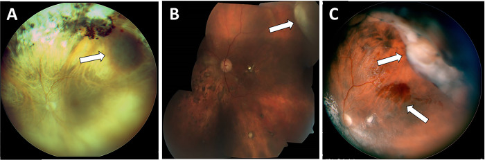
A is the hemorrhagic type, B the exudative type and C the combined type. The arrows point to the lesions.
The exudative type can look like vitreoretinal lymphoma, and the hemorrhagic type like melanoma.
Symptoms
Many patients (42%) have no symptoms, because the lesion lies far from the macula[1]. The lesion is found on a routine examination or on a widefield photograph. Others notice blurred vision, floaters from vitreous hemorrhage or a shadow in the visual field.
Vitreous hemorrhage seems to be more common in Asian patients. It occurred in 42% of eyes in Korean patients[2].
Imaging
Ultrasound
B-scan ultrasound shows a dome- or plateau-shaped elevation. How bright the inside of the lesion is (internal reflectivity, seen as the height of the spikes on the A-scan) varies. It was low in 41%, intermediate in 36% and high in 19%[1].
In about half of the eyes, a dark gap separates the clot from the choroid under it. This clot retraction cleft forms as the clot shrinks.
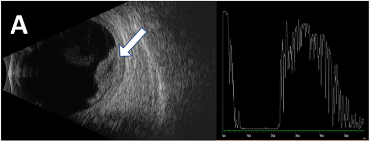
The left half is the B-scan, and the arrow points to the dome. The right half is the A-scan through the lesion, with high spikes inside it.
OCT
OCT shows what the dome is made of. In PEHCR the PED is often filled with blood. Subretinal fluid and blood may lie next to it.
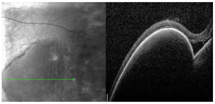
The green line on the left shows where the scan was taken. On the right, a tall PED lifts the retina, with a little subretinal fluid at its edge.
A standard OCT does not reach the far periphery. Widefield OCT helps when the device has it. Always scan the macula as well, to look for fluid, blood, drusen or cystoid macular edema.
Fluorescein angiography
On fluorescein angiography (FA), blood blocks the fluorescence and the lesion looks dark. Around it, areas of RPE atrophy look bright because the background fluorescence shows through (window defects), and exudative areas may leak. FA shows new vessels in only a minority of eyes.
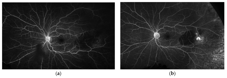
a: early phase. The hemorrhagic PED stays dark. b: late phase. The PED is still dark, and the lesion at its temporal edge leaks.
ICGA
ICGA sees through blood better than FA and shows the choroidal vessels. In a series of 48 eyes studied with widefield ICGA, 69% showed polypoidal lesions and 50% showed an abnormal choroidal vascular network in the area of the lesion[7]. Thick blood can still hide them. They may appear only in the late frames, and sometimes only on a later angiogram, after the blood clears.
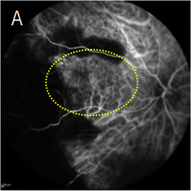
The dotted oval surrounds a branching network of abnormal choroidal vessels in the periphery.
Telling PEHCR from choroidal melanoma
Many patients with PEHCR are first referred as melanoma. In Korean patients, more than half were first diagnosed with choroidal melanoma, and only 8% were referred with the correct diagnosis[2].
A study that compared 40 eyes with PEHCR and 40 eyes with melanoma found features that appeared in only one of the two groups[8].
- Seen only in the PEHCR group: a hemorrhagic PED, a clot retraction cleft on ultrasound, lipid exudate and lesions in both eyes.
- Seen only in the melanoma group: orange pigment on the surface, and on ultrasound, choroidal excavation, a mushroom shape and extension into the ciliary body.
Every eye with PEHCR had at least one of its four features, and a hemorrhagic PED was the most useful single sign. These are not absolute rules: 3% of eyes with PEHCR showed choroidal excavation[1]. Melanoma also tends to show orbital shadowing on ultrasound and its own tumor vessels (a dual circulation) on FA[9]. When doubt remains, MRI with contrast can help: a melanoma enhances, while blood under the RPE does not.
Other lesions to consider are a ruptured retinal arterial macroaneurysm, a vasoproliferative tumor, choroidal metastasis and, for the exudative type, vitreoretinal lymphoma.
Course and treatment
Most lesions settle on their own
Most lesions (89%) stayed stable or shrank over a mean of 15 months[1]. The blood is absorbed and leaves RPE atrophy, pigment or a flat scar.
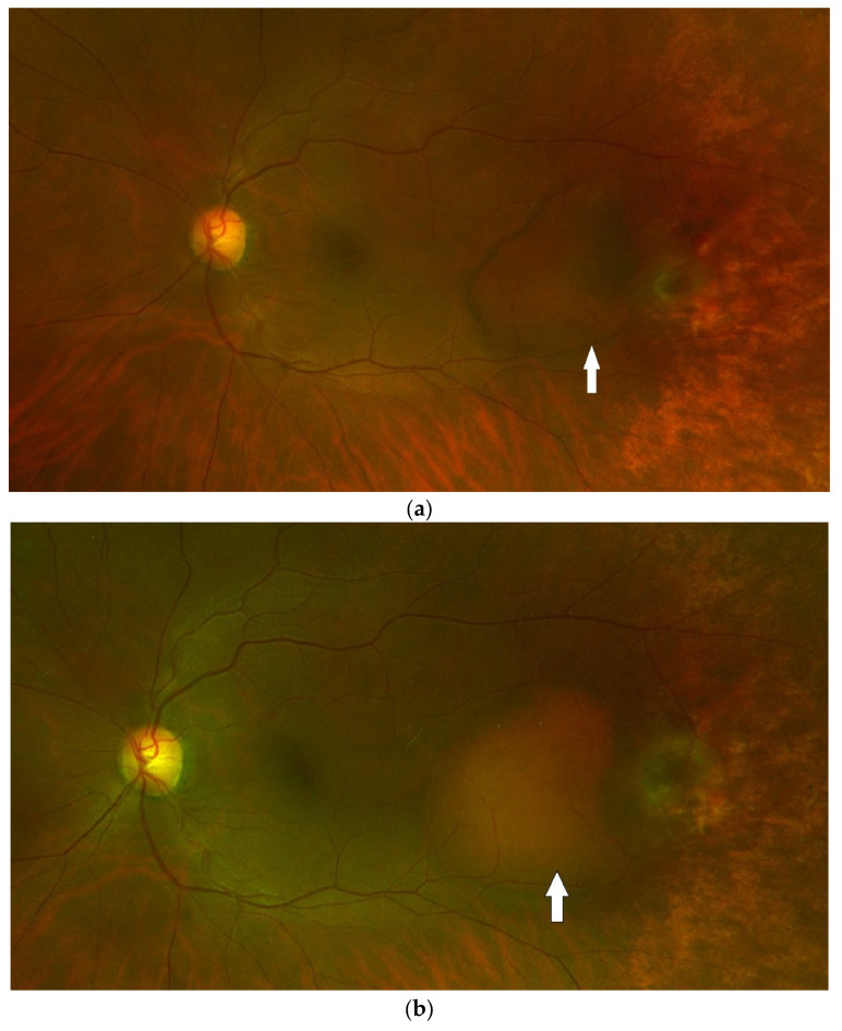
a: at presentation, a large PED (arrow) with blood around it. b: three months later without treatment, the blood has cleared.
An asymptomatic lesion far from the macula can be watched. Ask the patient to return early if new floaters or blurred vision appear.
Who needs treatment
Treat when the lesion threatens vision. The risk is higher when the lesion is large or has subretinal fluid next to it, and in these eyes spread to the macula was less common after anti-VEGF[10].
When fluid or blood moves toward the macula, intravitreal anti-vascular endothelial growth factor (anti-VEGF) injections are the usual treatment. About three injections dried the macula, but the fluid came back in some eyes[11].
When vitreous hemorrhage does not clear, do a vitrectomy. In a series of 84 eyes, it was the only treatment that improved vision[12]. It also lets you see the lesion when a tumor has not been ruled out.
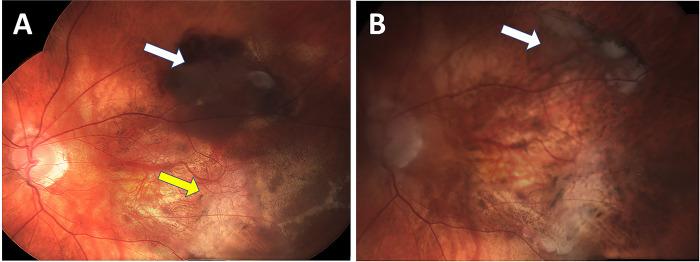
A: before treatment. The white arrow points to a hemorrhagic lesion at the superior arcade. The yellow arrow points to an old fibrotic lesion. B: after 10 injections, the blood has gone, and atrophy is left (white arrow).
The evidence comes from small retrospective series, with no randomized trial. Bevacizumab cleared the lesion in 8 of 9 eyes, but final vision did not differ from untreated eyes[6]. Laser, photodynamic therapy and cryotherapy have also been used, but in a Korean report one eye after laser and one after cryotherapy later bled massively under the fovea[13].
Summary
- PEHCR is blood, exudate and PED in the temporal periphery of older adults.
- It is often referred as melanoma. A hemorrhagic PED, lipid exudate or a clot retraction cleft points to PEHCR.
- ICGA often shows polypoidal lesions and an abnormal vessel network, like PCV.
- Most lesions settle on their own. Anti-VEGF is for macular threat, and vitrectomy for vitreous hemorrhage that does not clear.
References
[1] Shields CL, Salazar PF, Mashayekhi A, Shields JA. Peripheral exudative hemorrhagic chorioretinopathy simulating choroidal melanoma in 173 eyes. Ophthalmology. 2009;116(3):529-535. PMID 19157563.
[2] Lee H, Choi YJ, Lee SM, et al. Clinical characteristics of peripheral exudative hemorrhagic chorioretinopathy easily misdiagnosed at initial presentation in Korean patients. Sci Rep. 2025;15:30693. PMID 40841816.
[3] Mai S, Chen J, Li B, Sun X, Yu X. Peripheral exudative hemorrhagic chorioretinopathy: a comprehensive review of clinical features, differential diagnosis, pathogenesis-related disease spectra, and management. Surv Ophthalmol. 2026 (online ahead of print). PMID 42906862.
[4] Goldman DR, Freund KB, McCannel CA, Sarraf D. Peripheral polypoidal choroidal vasculopathy as a cause of peripheral exudative hemorrhagic chorioretinopathy: a report of 10 eyes. Retina. 2013;33(1):48-55. PMID 22836900.
[5] Mantel I, Uffer S, Zografos L. Peripheral exudative hemorrhagic chorioretinopathy: a clinical, angiographic, and histologic study. Am J Ophthalmol. 2009;148(6):932-938. PMID 19800613.
[6] Safir M, Zloto O, Fabian ID, et al. Peripheral exudative hemorrhagic chorioretinopathy with and without treatment—clinical and multimodal imaging characteristics and prognosis. PLoS One. 2022;17(9):e0275163. PMID 36166419.
[7] Mantel I, Schalenbourg A, Zografos L. Peripheral exudative hemorrhagic chorioretinopathy: polypoidal choroidal vasculopathy and hemodynamic modifications. Am J Ophthalmol. 2012;153(5):910-922. PMID 22310077.
[8] Sodhi GS, Singh N, Wrenn J, Singh AD. Peripheral hemorrhagic chorioretinopathy: differentiating features from choroidal melanoma. Ocul Oncol Pathol. 2023;9(1-2):1-8. PMID 37600851.
[9] Salmon JF. Kanski’s Clinical Ophthalmology: A Systematic Approach. 10th ed. Elsevier; 2025. Chapter 20, Ocular tumours: Choroidal melanoma.
[10] Zicarelli F, Preziosa C, Staurenghi G, Pellegrini M. Peripheral exudative haemorrhagic chorioretinopathy: a widefield imaging study. Br J Ophthalmol. 2021;105(10):1410-1414. PMID 32883656.
[11] Seibel I, Hager A, Duncker T, et al. Anti-VEGF therapy in symptomatic peripheral exudative hemorrhagic chorioretinopathy (PEHCR) involving the macula. Graefes Arch Clin Exp Ophthalmol. 2016;254(4):653-659. PMID 26148802.
[12] Vandefonteyne S, Caujolle JP, Rosier L, et al. Diagnosis and treatment of peripheral exudative haemorrhagic chorioretinopathy. Br J Ophthalmol. 2020;104(6):874-878. PMID 31645320.
[13] Kim YT, Kang SW, Lee JH, Chung SE. Peripheral exudative hemorrhagic chorioretinopathy in Korean patients. Jpn J Ophthalmol. 2010;54(3):227-231. PMID 20577857.
Which retinal layer makes which wave, the three abnormal patterns, and the dark-adaptation trap to check before you report “negative” — on one page, by email. One email when I publish something new, about once a month. Unsubscribe anytime.