NMOSD vs MOGAD Optic Neuritis: Differences and Treatment
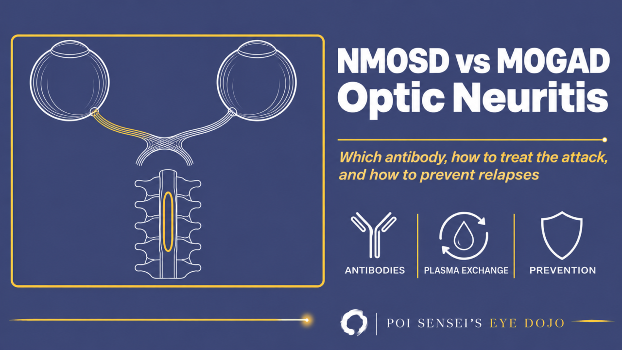
A patient with optic neuritis has lost all light perception in the affected eye, and steroid pulses do not help. That picture should make you think of neuromyelitis optica spectrum disorder (NMOSD) with antibodies to aquaporin-4 (AQP4). Its partner is MOG antibody-associated disease (MOGAD), and the MOG antibody is the other test you send in every optic neuritis.
This article goes through how AQP4-positive optic neuritis differs from typical optic neuritis, the kind seen with multiple sclerosis (MS), and from MOGAD, how the attack is treated, and which drugs prevent relapses.
AQP4-positive optic neuritis
AQP4 is a water channel found in large amounts on the foot processes of astrocytes. Antibodies against it bind there, switch on complement and destroy the astrocytes, causing severe inflammation of the optic nerve and spinal cord.
The disease used to be called neuromyelitis optica (NMO), or Devic disease. Under the 2015 international criteria it is now called NMOSD, and patients with AQP4 antibodies make up most of it.
AQP4-positive optic neuritis is an acute optic neuritis of one or both eyes, seen mostly in middle-aged women. Women outnumber men by up to 10 to 1, and onset peaks around age 40, although it can start in childhood or in old age[1]. In a Japanese nationwide study of optic neuritis, 12% of patients were AQP4-antibody positive, and their median age at onset was 53[2].
How it differs from typical optic neuritis
Compared with typical optic neuritis, AQP4-positive optic neuritis has these features:
- Vision loss is often severe, down to hand motions or no light perception
- It tends to relapse, and the visual prognosis is poor
- It often takes the form of retrobulbar optic neuritis, without disc swelling
The field defect is not always a central scotoma either. It can be altitudinal, as in ischemic optic neuropathy, or bitemporal when the inflammation reaches the optic chiasm. Defects other than a central scotoma are seen in about a quarter of attacks[1].
Compared with MOGAD and MS
MOGAD affects men and women about equally and often starts in childhood or young adulthood. Vision at the worst point is often worse than 6/60 (20/200), but it often comes back quickly with steroids[3].
| AQP4+ | MOGAD | MS | |
|---|---|---|---|
| Age | Peak about 40 | Children, young adults | About 30 |
| Sex | Mostly women | Equal | More women |
| Vision loss | Severe | Severe, recovers well | Milder |
| Disc swelling | Uncommon | Common | Rare |
| Spinal MRI | Long lesion, 3+ segments | Long lesions, often the conus | Short lesions |
| Brain MRI | Often not MS-like | Can look like ADEM | Plaques by the ventricles |
| Steroid pulses | May not work | Work, but relapse on taper | Speed recovery |
ADEM is acute disseminated encephalomyelitis. The conus is the lower tip of the spinal cord.
MS drugs can make NMOSD worse. Interferon beta, natalizumab, fingolimod, alemtuzumab and dimethyl fumarate have all been reported to trigger severe attacks in NMOSD[4]. Telling these diseases apart matters for the treatment as much as for the diagnosis.
Treating the attack
All three are usually treated first with high-dose intravenous methylprednisolone (IVMP), or steroid pulse therapy. In AQP4-positive disease, if the steroid pulses do not work, the next step is plasma exchange.
Do not wait too long. In a severe attack, plasma exchange can also be started together with the steroids. When plasma exchange or a similar procedure began within 2 days of onset, up to 40% of patients recovered completely, against 3.7% when it began 7 days or later[4]. So when the clinical picture strongly suggests AQP4-positive disease, I start preparing plasma exchange right away, without waiting for the antibody result.
In my own practice, a patient with no light perception who did not respond to steroid pulses recovered to a corrected acuity of about 20/30 with plasma exchange. Even when steroids have failed, plasma exchange is still worth trying.
Do not keep repeating steroid pulses that are not working.
Preventing relapses in AQP4-positive NMOSD
Almost every AQP4-positive patient relapses, so long-term immunotherapy is started after the first attack. Oral steroids are continued after the attack as well.
I usually start prednisolone at 30 mg/day and taper it, going especially slowly around 15 mg/day, where relapses become more likely. The recommendations of the Neuromyelitis Optica Study Group (NEMOS), a German expert network, taper to 10–15 mg/day within 2–3 weeks and continue a low dose for up to 3–6 months. In small studies from Asia, attacks were more likely with fast tapering and with doses below 10 mg/day[4].
Azathioprine or mycophenolate mofetil can be added to the steroids and has been reported to work well, although neither is approved for NMOSD.
Since 2019, biologic drugs have been approved for AQP4-positive NMOSD:
| Drug | Target | US approval |
|---|---|---|
| Eculizumab | Complement C5 | Yes |
| Ravulizumab | C5 (long-acting) | Yes |
| Inebilizumab | CD19 | Yes |
| Satralizumab | IL-6 receptor | Yes |
| Rituximab | CD20 | No (yes in Japan) |
Rituximab is widely used off-label in the US and Europe. All four approved drugs are licensed only for AQP4-positive disease, not for MOGAD.
The figure shows where each drug acts in the disease process.
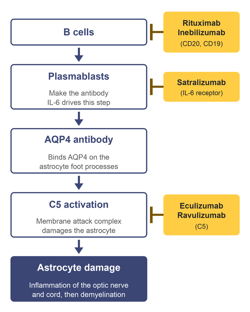
The left column is the disease process, from top to bottom. B cells become plasmablasts and make the AQP4 antibody. The antibody binds AQP4 on astrocytes and activates complement, which destroys the astrocytes. Inflammation of the optic nerve and spinal cord follows, with demyelination as a secondary step.
The yellow boxes on the right are the drugs, and the ⊥ at the end of each line is that drug’s main point of action. Rituximab depletes CD20-positive B cells, and inebilizumab depletes CD19-positive B cells and plasmablasts. Satralizumab blocks the IL-6 receptor and reduces plasmablast differentiation and antibody secretion. Eculizumab and ravulizumab block complement C5 and reduce the complement damage to astrocytes.
Other autoimmune diseases
Patients with AQP4-positive NMOSD often carry other autoantibodies too, such as antinuclear antibodies, rheumatoid factor, SS-A antibodies, thyroid antibodies and acetylcholine receptor antibodies. Signs of another autoimmune disease, such as lupus, Sjögren syndrome or myasthenia gravis, are reported in 25–40% of patients[1].
In my experience, patients with a connective tissue disease relapse more and are harder to control. Before the newer relapse-prevention drugs came out, I followed a patient with lupus who relapsed while taking prednisolone 30 mg/day and needed azathioprine added.
The disease can take a little more vision with each relapse and end in blindness. Keeping up relapse prevention matters more than anything.
Summary
- Severe optic neuritis with poor vision or a poor steroid response suggests AQP4-positive NMOSD.
- MOGAD often swells the disc and responds to steroids, but can relapse on the taper.
- MS drugs can make NMOSD worse.
- Start with steroid pulses, and move to plasma exchange early if they do not work.
- Relapse prevention is the key: oral steroids, immunosuppressants and four approved biologics.
References
[1] Jarius S, Aktas O, Ayzenberg I, et al. Update on the diagnosis and treatment of neuromyelits optica spectrum disorders (NMOSD) – revised recommendations of the Neuromyelitis Optica Study Group (NEMOS). Part I: Diagnosis and differential diagnosis. J Neurol. 2023;270(7):3341–3368. PMID 37022481.
[2] Ishikawa H, Kezuka T, Shikishima K, et al. Epidemiologic and Clinical Characteristics of Optic Neuritis in Japan. Ophthalmology. 2019;126(10):1385–1398. PMID 31196727.
[3] Banwell B, Bennett JL, Marignier R, et al. Diagnosis of myelin oligodendrocyte glycoprotein antibody-associated disease: International MOGAD Panel proposed criteria. Lancet Neurol. 2023;22(3):268–282. PMID 36706773.
[4] Kümpfel T, Giglhuber K, Aktas O, et al. Update on the diagnosis and treatment of neuromyelitis optica spectrum disorders (NMOSD) – revised recommendations of the Neuromyelitis Optica Study Group (NEMOS). Part II: Attack therapy and long-term management. J Neurol. 2024;271(1):141–176. PMID 37676297.
Which retinal layer makes which wave, the three abnormal patterns, and the dark-adaptation trap to check before you report “negative” — on one page, by email. One email when I publish something new, about once a month. Unsubscribe anytime.