Corneal Dystrophies: How to Tell Them Apart by Layer
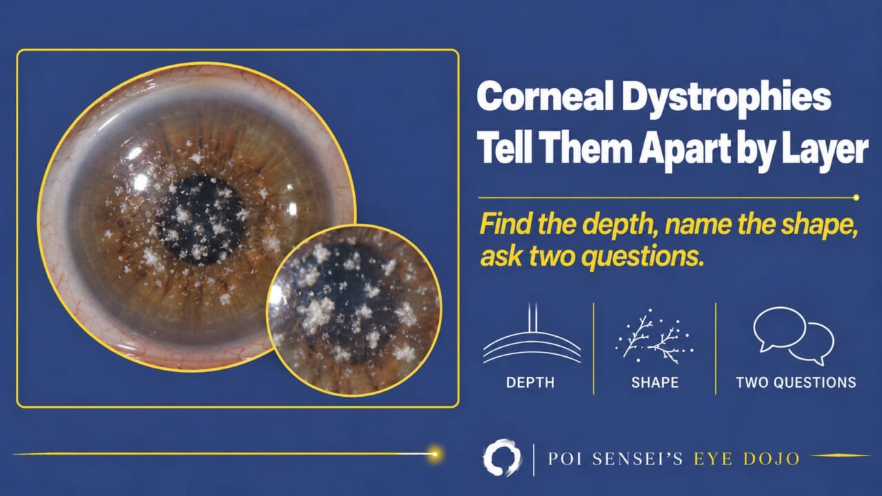
There are more than twenty corneal dystrophies, most of them named after people, and several have been renamed in the last ten years. You do not need to memorize the list. Each dystrophy sits at a particular depth, and the depth, the shape of the opacity and two questions to the patient narrow it down to a handful. This article goes through the cornea from front to back, using the four groups of the current international classification, IC3D edition 3 (2024)[1].
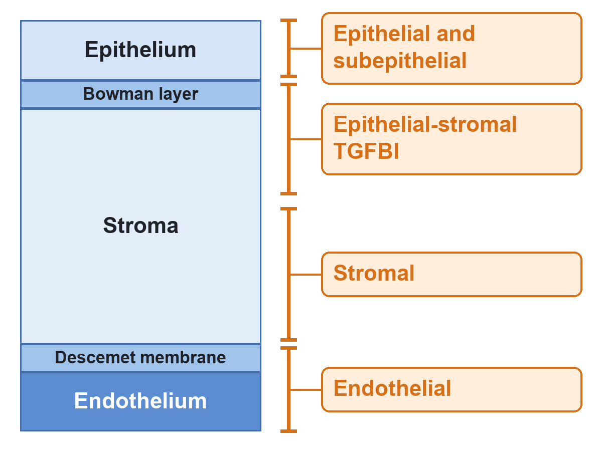
- Find the depth. A thin optical section shows whether the change sits in the epithelium, at Bowman layer and the anterior stroma, within the stroma, or at Descemet membrane and the endothelium.
- Name the shape: maps and dots, tiny cysts, branching lines, crumbs, flecks with haze between them, crystals, guttae, vesicles or bands.
- Ask two questions: Have you had attacks of pain on waking? and Does anyone in your family have the same thing?
- Then place it in one of the four IC3D groups below.
The four groups, and why "Bowman layer dystrophies" disappeared
The IC3D (International Committee for Classification of Corneal Dystrophies) groups the dystrophies anatomically: epithelial and subepithelial, epithelial-stromal TGFBI, stromal and endothelial[1]. Older textbooks had a fifth group, the Bowman layer dystrophies. Since the second edition in 2015, those dystrophies, together with lattice and granular dystrophy, have been gathered under the one gene they share, TGFBI, because they also share inheritance, histology and a tendency to recurrent erosions even though they look different at the slit lamp[1].
Many textbooks, including the current edition of Kanski, still use the older arrangement with a separate Bowman layer group[2]. The two map onto each other easily, and the general rule holds in both: a dystrophy is bilateral, affects mainly one layer, and progresses slowly[2]. That is why depth is the first question.
Epithelium and subepithelium
Epithelial basement membrane dystrophy (EBMD)
EBMD is the one you will see most often. Strictly speaking it is hardly a dystrophy: most cases have no family history, and IC3D regards it as probably degenerative[1]. Look for gray maps with scalloped edges, putty-gray dots clustered like an archipelago, and parallel curved fingerprint lines[1]. Direct illumination can miss them. Retroillumination or scleral scatter brings them out[1][2]. Because the abnormal epithelium is slightly raised, fluorescein flows around it and leaves a dark patch of negative staining[1]. The findings change over time and may be absent or subtle in the fellow eye[2].
The presenting complaint is recurrent erosion: sharp pain on waking, when the lid pulls the loosely attached epithelium off. Only about one in ten patients ever has erosions, usually in their twenties[2]. Bilateral erosions with no history of trauma should make you look for this dystrophy[2].
Treatment is lubricants by day and ointment at night, a bandage lens for an attack, and for persistent cases debridement or phototherapeutic keratectomy (PTK)[1]. In a refractive surgery candidate, EBMD favors surface ablation over LASIK, which can loosen the epithelium[3].
Meesmann corneal dystrophy
Meesmann corneal dystrophy is autosomal dominant (the keratin genes KRT3 and KRT12) and shows countless tiny intraepithelial cysts, densest in the center and spreading toward the periphery[1][2]. On direct light they read as a faint gray haze. On indirect light each cyst becomes a clear bubble. Most patients have little more than irritation or glare, and treatment is rarely more than lubricants[1].
Gelatinous drop-like corneal dystrophy (GDLD)
GDLD is the odd one out in this group: autosomal recessive (TACSTD2), with onset in the first two decades[1]. Subepithelial amyloid forms mulberry-like nodules, or early on a band that looks like band keratopathy[1]. The epithelium is hyperpermeable, so the lesions stain with fluorescein, superficial vessels grow in, and vision falls steadily[1].
A bandage contact lens slows the growth of the amyloid nodules. Once the nodules get in the way of the lens, remove them with a superficial keratectomy[1]. It recurs after superficial keratectomy and after keratoplasty, usually within a few years[1]. It is rare in most countries. In Japan, where I practice, it is one of the dystrophies you are expected to recognize.
Bowman layer and anterior stroma: the TGFBI group
Five autosomal dominant dystrophies map to TGFBI on chromosome 5. All five can cause recurrent erosions, and all five can recur in a graft[1]. The shape of the deposit tells them apart.
Classic lattice corneal dystrophy
Classic lattice corneal dystrophy has thin, branching, refractile lines that start centrally and superficially and spread outward and deeper, with a ground-glass subepithelial haze and whitish dots. The far periphery, Descemet membrane and the endothelium stay clear[1]. The deposit is amyloid, Congo red positive[1]. Erosions start early, often before the lines themselves are visible, and vision is usually poor by about age 40[1][2]. Retroillumination is the way to see the lines. Lattice variants (other TGFBI mutations) have thicker, deeper lines and start later[1].
Granular corneal dystrophy type 1
Granular corneal dystrophy type 1 gives discrete, well-defined gray-white granules with clear stroma between them, like crumbs. They never reach the limbus, and over the years they coalesce into snowflakes and sink deeper[1]. The deposit is keratohyalin, Masson trichrome positive[1]. Glare and photophobia come first, erosions and visual loss later[1].
Granular corneal dystrophy type 2 (formerly Avellino dystrophy)
Granular corneal dystrophy type 2 is a mixture of granular type 1 and lattice: superficial whitish rings and discs, plus star-shaped deposits deeper in the stroma[1]. Histology shows both keratohyalin and amyloid, so both Masson trichrome and Congo red stain[1]. Erosions and visual loss are milder than in type 1[1].
Reis–Bücklers corneal dystrophy
Reis–Bücklers corneal dystrophy forms confluent, coarse, geographic gray opacities at the level of Bowman layer, from childhood, with painful erosions that may come before the opacities[1]. Keratohyalin again, red with Masson trichrome[1].
Thiel–Behnke corneal dystrophy
Thiel–Behnke corneal dystrophy makes a subepithelial honeycomb pattern that spares the periphery, with fewer erosions and later visual loss than Reis–Bücklers. In an individual patient the two can be impossible to separate without genetic testing[1].
Meretoja syndrome: no longer a corneal dystrophy
Meretoja syndrome, formerly called lattice corneal dystrophy type 2, is no longer in this group: its lattice lines come from systemic gelsolin amyloidosis (familial amyloidosis, Finnish type), and IC3D no longer counts it as a corneal dystrophy[1]. Its lines are sparse, start at the periphery and spread centrally, the reverse of classic lattice, erosions are rare, and the cornea is hypoesthetic, which matters if the patient ever needs a graft[1][2].
Look at the face as well: a cranial neuropathy gives mask-like facies and, at times, an exposed cornea[2][3].
Stroma
Macular corneal dystrophy
Macular corneal dystrophy looks like granular dystrophy at first glance. Three things separate them: it is autosomal recessive (CHST6), the stroma between the irregular whitish flecks is hazy rather than clear, and the opacities reach the limbus and go down to Descemet membrane[1]. Severe visual loss arrives between 10 and 30 years of age[1].
The deposit is glycosaminoglycan, stained by Alcian blue or colloidal iron, and the endothelium is involved: Descemet membrane thickens and guttae appear[1]. The cornea is thin early on and thickens only late, when the endothelium fails, and corneal sensation is reduced[2]. Because the endothelium is involved, a full-thickness (penetrating) graft may be better than a deep anterior lamellar one[1]. Recurrence in the graft is less common than in granular or lattice dystrophy[1].
Schnyder corneal dystrophy
Schnyder corneal dystrophy used to be called Schnyder crystalline corneal dystrophy. The word was dropped because only half of patients have crystals, and the other half were being missed[1]. It is autosomal dominant (UBIAD1). A central disc or ring, sometimes with crystals, appears in young adults. Arcus follows, and later the whole cornea becomes hazy[1].
The deposit is cholesterol and phospholipid, and the pathologist needs fresh tissue for Oil Red O, because routine processing dissolves it[1]. Vision is often better than the cornea suggests, but it drops in bright light, and glare is the main complaint[1]. Both affected and unaffected family members may have hyperlipoproteinemia[1].
Fleck, posterior amorphous, central cloudy and pre-Descemet
The remaining stromal entries, fleck, posterior amorphous, central cloudy and pre-Descemet corneal dystrophy, are usually found by chance and need no treatment[1].
Descemet membrane and endothelium
Fuchs endothelial corneal dystrophy (FECD)
FECD begins with guttae: small bumps on Descemet membrane that appear centrally and spread outward, and that give the endothelium a beaten-metal look in specular reflection, with or without pigment dusting[1]. That is stage 1. Some patients never move beyond it; others progress to stromal edema (stage 2) and then epithelial bullae and bullous keratopathy (stage 3)[1].
The symptom that gives it away is blur that is worst in the morning, because the stroma swells behind closed lids overnight and clears during the day[1]. Most cases begin in middle age or later, with a female predominance of about 2.5 to 3.5 to 1; the rare early-onset form (COL8A2) starts in the first two decades[1][2].
Guttae alone can leave excellent vision and need no treatment. Hypertonic saline drops and a hair dryer held at arm’s length in the morning buy time once mild edema appears[2]. Once edema reduces vision, endothelial keratoplasty (DMEK or DSAEK) is the standard. Penetrating keratoplasty is kept for end-stage scarring[1].
Cataract surgery costs endothelial cells, and a Fuchs cornea has few to spare. Persistent edema after cataract surgery is more likely when the preoperative central corneal thickness is above about 630 to 640 µm, and in an eye that already has edema a combined cataract and keratoplasty procedure can be considered[2]. Measure the central corneal thickness before you book the surgery.
In Japan, FECD is far less common than in Europe or North America, and the TCF4 repeat expansion that underlies most Western cases is found in only a minority of Japanese patients. So I see guttae far more often than I see Fuchs dystrophy.
Posterior polymorphous corneal dystrophy (PPCD)
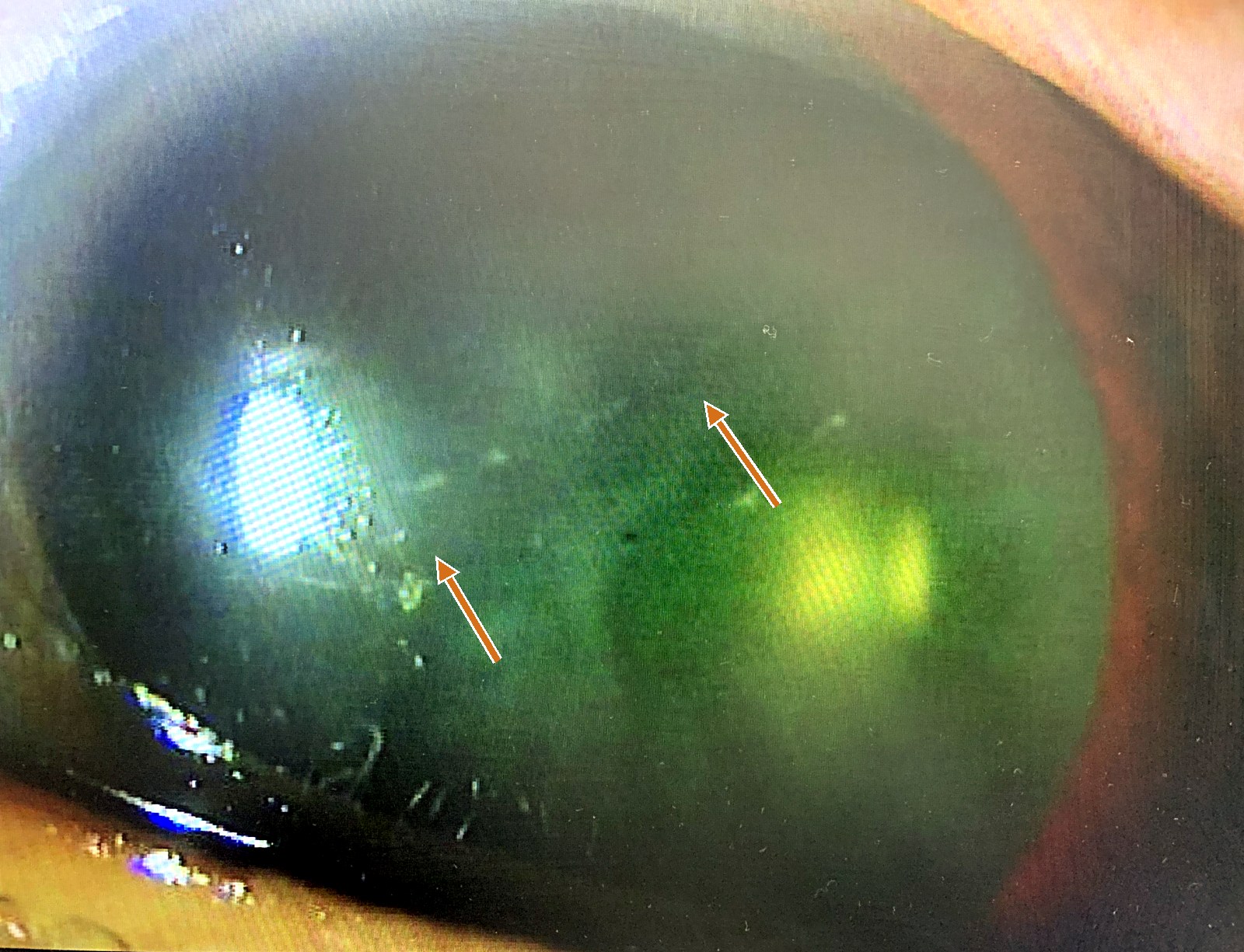
PPCD is autosomal dominant and often asymmetric, with several kinds of endothelial finding: grouped vesicles, parallel gray-white bands with flaky edges ("railroad tracks") that can cross the whole cornea, and geographic gray opacities of Descemet membrane[1]. Retroillumination shows them best[1].
Most patients are asymptomatic for life, but some develop corneal edema that needs a graft, peripheral iridocorneal adhesions or raised intraocular pressure[1]. So every patient with PPCD gets gonioscopy and a pressure check. An association with Alport syndrome has also been described[2]. The main differential is iridocorneal endothelial (ICE) syndrome, which is sporadic, usually unilateral and comes with iris atrophy.
Posterior corneal vesicle: the unilateral look-alike
Before you call a band or vesicle unilateral, look at the other eye with specular microscopy: a "unilateral" vesicle sometimes turns out to be PPCD with a quiet fellow eye. A truly unilateral band or vesicle with the same appearance, no family history and no progression is what I call a posterior corneal vesicle. IC3D lists these as isolated unilateral cases with a similar phenotype but no known heredity[1]. Two things help me separate it from a Descemet tear after forceps delivery: the history, and the direction. Birth-trauma tears usually run vertically; posterior corneal vesicles usually run horizontally.
Congenital hereditary endothelial dystrophy (CHED)
CHED is autosomal recessive (SLC4A11): a cloudy, thickened cornea from birth, often with nystagmus, little progression, and keratoplasty when vision demands it[1]. Vision is sometimes better than the cornea leads you to expect, so measure it before you decide[2]. If you learned an autosomal dominant "CHED1", it has gone: the families described under that name most likely had PPCD, and IC3D removed it in 2015[1].
- Autosomal recessive: macular, gelatinous drop-like, CHED. Everything else in this article is dominant or sporadic.
- Painful recurrent erosions: EBMD, Reis–Bücklers, Thiel–Behnke, classic lattice, granular type 1 (type 2 less so).
- What the deposit is: amyloid (Congo red) in lattice, gelatinous drop-like and granular type 2; keratohyalin (Masson trichrome) in Reis–Bücklers and granular types 1 and 2; glycosaminoglycan (Alcian blue, colloidal iron) in macular; lipid (Oil Red O) in Schnyder.
- Endothelium involved outside the endothelial group: macular corneal dystrophy, which is why its graft is often a penetrating one.
Summary
- Find the depth first, then name the shape. Then ask about pain on waking and family history.
- Epithelium: maps, dots and fingerprints are EBMD; a sheet of tiny cysts is Meesmann; mulberry nodules in a young patient are gelatinous drop-like dystrophy.
- TGFBI group: lattice lines, crumbs with clear stroma between, a mixture of rings and stars, or geographic or honeycomb opacities at Bowman layer. The mixture (granular type 2) rules out refractive surgery.
- Stroma: flecks with diffuse haze reaching the limbus are macular dystrophy; a central disc with or without crystals is Schnyder.
- Endothelium: guttae with morning blur are Fuchs; vesicles and railroad tracks are PPCD (check the angle and pressure); a cloudy cornea from birth is CHED.
References
[1] Weiss JS, Rapuano CJ, Seitz B, et al. IC3D classification of corneal dystrophies—edition 3. Cornea. 2024;43(4):466–527. PMID 38359414.
[2] Salmon JF. Kanski’s Clinical Ophthalmology: A Systematic Approach. 10th ed. Elsevier; 2025. Chapter 7, Cornea: corneal dystrophy.
[3] Yanoff M, Duker JS, eds. Ophthalmology. 5th ed. Elsevier; 2019. Chapters 4.19–4.21: anterior and stromal corneal dystrophies, diseases of the corneal endothelium.