Choroideremia: Fundus, OCT and ERG Findings
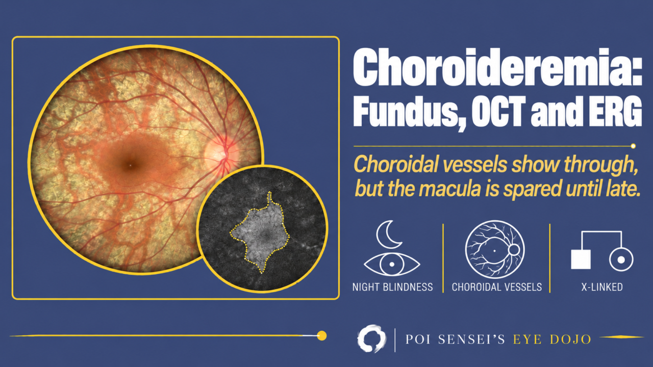
A boy has trouble seeing in the dark. Years later, his fundus shows large choroidal vessels over a pale background, while the macula still looks normal. Think of choroideremia. This article explains its inheritance and symptoms, how to read the fundus, OCT and ERG, and how to tell it from retinitis pigmentosa.
- A boy or man has night blindness from childhood.
- The retina and choroid are atrophic, and the choroidal vessels show through.
- The macula is spared until late.
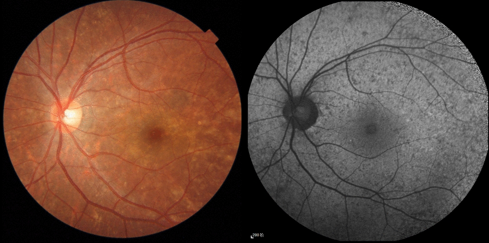
- Female relatives may have a mottled fundus.
What is choroideremia?
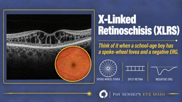
Choroideremia is an inherited retinal disease with X-linked recessive inheritance. It mainly affects males, but female carriers may also show a mild, mottled fundus. Another X-linked recessive retinal disease is X-linked retinoschisis.
Symptoms
The typical patient notices night blindness in childhood.
The visual field first shows a ring scotoma, and then constricts concentrically. Central vision usually stays good until about age 40, and then it falls. Visual acuity of 20/40 (6/12) is often kept into the 50s[1]. Severe vision loss usually comes after that, and some patients lose light perception[2].
Examination findings
Fundus
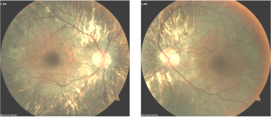
The retina and the choroid are severely atrophic, and the choroidal vessels show through. The atrophy starts in the mid-periphery and spreads toward the center. The macula is spared until late. In the end stage, only the large choroidal vessels remain over the white sclera[2].
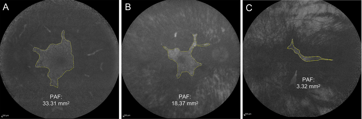
On fundus autofluorescence, areas where the retinal pigment epithelium (RPE) has been lost are dark (hypoautofluorescent). At the center, an area of normal autofluorescence remains. This area becomes smaller with age.
OCT
OCT shows loss of the RPE over a wide area and a thin choroid.
ERG
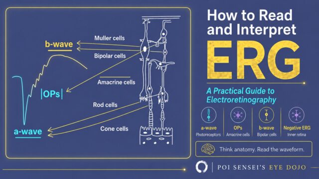
The ERG is reduced from early in the disease and often becomes flat. The rod response is lost first. The cone response may still be recordable, but it is severely reduced[2].
Differential diagnosis
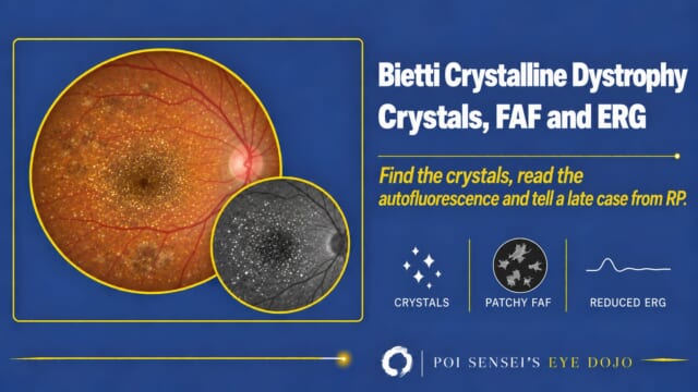
Retinitis pigmentosa with severe chorioretinal atrophy can be hard to tell apart from choroideremia. Genetic testing then makes the diagnosis: choroideremia is caused by variants in the CHM gene. Some families are first diagnosed with retinitis pigmentosa, and the diagnosis is corrected only after genetic testing[3].
Gyrate atrophy can also look similar. It is autosomal recessive, shows sharply demarcated round patches of atrophy, and raises the plasma ornithine level[2].
Treatment
There is no approved treatment. Gene therapy has been tested, but a phase 3 trial did not meet its main goal of improving visual acuity[4].
Summary
- Choroideremia is X-linked recessive. Males are affected, and female carriers may show a mild, mottled fundus.
- Night blindness from childhood, then a ring scotoma and a constricting visual field.
- Fundus: severe chorioretinal atrophy with visible choroidal vessels. The macula is spared until late.
- ERG: severely reduced and often flat. Genetic testing for CHM separates it from retinitis pigmentosa.
Figure credits
Fundus photographs: Figure 2 from Guo H, Li J, Gao F, et al. BMC Ophthalmol 2015;15:85 (https://doi.org/10.1186/s12886-015-0081-4), licensed under CC BY 4.0. The lower row (patient IV-5) cropped, without the row label.
Fundus autofluorescence: Figure 5 from Hayashi T, Kameya S, Mizobuchi K, et al. Sci Rep 2020;10(1):15883 (https://doi.org/10.1038/s41598-020-72623-1), licensed under CC BY 4.0. Panels A to C rearranged in one row, without panel D.
Female carrier: Figure 2 from Han X, Yu Y, Ding J, et al. Eye Vis (Lond) 2026;13(1):9 (https://doi.org/10.1186/s40662-026-00476-2), licensed under CC BY 4.0. Column a cropped, with the fundus photograph and the autofluorescence image placed side by side.
References
[1] Hayashi T, Kameya S, Mizobuchi K, et al. Genetic defects of CHM and visual acuity outcome in 24 choroideremia patients from 16 Japanese families. Sci Rep. 2020;10(1):15883. PMID 32985515.
[2] Salmon JF. Kanski’s Clinical Ophthalmology: A Systematic Approach. 10th ed. Elsevier; 2025. Chapter 15, Inherited retinal degenerations: generalized choroidal dystrophies.
[3] Guo H, Li J, Gao F, et al. Whole-exome sequencing reveals a novel CHM gene mutation in a family with choroideremia initially diagnosed as retinitis pigmentosa. BMC Ophthalmol. 2015;15:85. PMID 26216097.
[4] MacLaren RE, Fischer MD, Gow JA, et al. Subretinal timrepigene emparvovec in adult men with choroideremia: a randomized phase 3 trial. Nat Med. 2023;29(10):2464-2472. PMID 37814062.
Which retinal layer makes which wave, the three abnormal patterns, and the dark-adaptation trap to check before you report “negative” — on one page, by email. One email when I publish something new, about once a month. Unsubscribe anytime.