Leber Congenital Amaurosis: How to Recognize It in an Infant
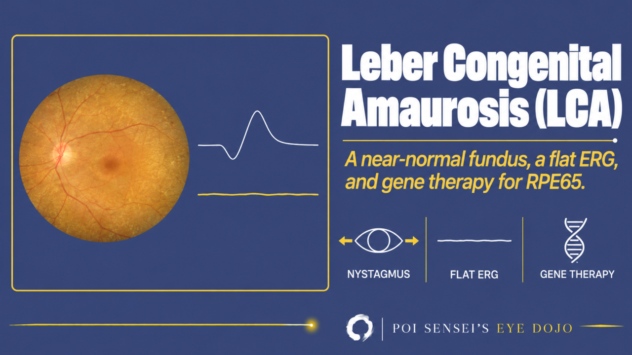
A baby does not follow faces or lights, has nystagmus, and the fundus looks almost normal. Think of Leber congenital amaurosis (LCA). This article shows how to recognize LCA in an infant, why the electroretinogram (ERG) is the key test, and which form can now be treated.
- Severe visual loss in both eyes is noticed in the first year of life.
- There is nystagmus or roving eye movements, and the pupils react poorly to light.
- The child presses, rubs or pokes the eyes.
- The fundus looks normal or nearly normal, but the ERG is flat (almost no response) or severely reduced.
What is Leber congenital amaurosis?
LCA is also called congenital amaurosis. "Amaurosis" means loss of vision in an eye that looks normal from the outside.
LCA is a group of inherited retinal dystrophies. Vision is very poor from birth or from the first few months of life. It is rare, affecting about 1 in 33,000 to 81,000 people[1].
Symptoms
The poor vision is usually noticed at birth or within the first year. The baby does not follow light or faces, the eyes wander or show nystagmus, and the pupil reactions are weak.
Children with severe visual loss often press, rub or poke their eyes. This is the oculodigital sign. Rubbing the eyes makes the child see flashes of light (phosphenes), so these children keep doing it.
Constant rubbing can shrink the fat in the orbit, so the eyes sink back (enophthalmos). Rubbing can also lead to keratoconus[2].
Many affected infants have high hyperopia. Most are otherwise healthy. If the child also has developmental delay or kidney problems, think of a syndrome such as Joubert or Senior-Løken syndrome[1].
Rods first or cones first?
In most forms of LCA, the rods are damaged first and the cones later. In some forms, the cones are damaged first. The two groups look different in clinic.
Cones first
GUCY2D is one of the most common causes of LCA, and it causes this type. Vision is very poor from birth, and the child is often strongly bothered by light. The fundus often looks nearly normal. These children often keep part of their rod function.
Rods first
RPE65 causes this type. Vision in dim light is poor from birth, but some daylight vision from the cones remains in early childhood. Nystagmus is often mild. Many of these children have the milder form, called early-onset severe retinal dystrophy (EOSRD, below). RPE65 disease is the form that gene therapy can treat[1].
Inheritance and fundus
Most LCA is autosomal recessive, but a few genes cause autosomal dominant disease. At least 28 genes are known, and the findings differ from gene to gene.
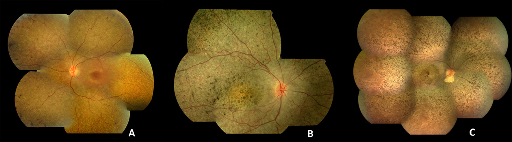
The fundus may be normal in early life, apart from mildly narrow arteries. With age, pigment changes, macular atrophy, optic atrophy and very narrow arteries appear[2].
On OCT, when the macula is atrophic, the ellipsoid zone (EZ), the line made by the photoreceptors, is often unclear or lost.
ERG
The ERG is flat or severely reduced from early on, even when the fundus looks normal[3]. This is the key test.
In some forms, a small response may remain from the photoreceptors that are less damaged. For example, children with GUCY2D-LCA often keep part of their rod function[1].
Telling LCA from other causes
Several conditions also cause nystagmus and poor vision in infants. The ERG helps separate them.
| Condition | ERG |
|---|---|
| LCA | Flat or severely reduced |
| Achromatopsia | No cone response, normal rods |
| Congenital stationary night blindness | Usually b-wave smaller than a-wave |
| Albinism | Normal |
| Optic nerve hypoplasia | Normal |
Achromatopsia and congenital stationary night blindness do not progress. LCA usually does[1].
Early-onset severe retinal dystrophy
Some children develop night blindness or poor vision later, after infancy and usually before age 5. This is called early-onset severe retinal dystrophy (EOSRD). Vision is better than in LCA, and small ERG responses may remain. EOSRD shares its genes with LCA, so the two are seen as one spectrum[1].
Treatment
For most forms of LCA, there is no treatment that stops the disease. For LCA caused by mutations in both copies of RPE65, gene therapy with voretigene neparvovec (Luxturna) is approved in the US (2017), the EU (2018) and Japan (2023)[4]. It is a single injection under the retina.
Genetic testing finds the children who may benefit from gene therapy or clinical trials. It also shows which children need further checks for systemic disease[1].
Low-vision aids, advice on schooling and rehabilitation also help.
Summary
- LCA is an inherited retinal dystrophy with severe visual loss from birth, nystagmus, weak pupil responses and the oculodigital sign.
- Most forms damage the rods first. In the cones-first forms, vision is very poor from birth.
- The fundus can look normal early. The ERG is flat or severely reduced, and it is the key test.
- EOSRD starts later and is milder, on the same spectrum as LCA.
- RPE65-LCA can be treated with gene therapy, so send every child for genetic testing.
Figure credits
Fundus photographs: Figure 2 from Kumaran N, Moore AT, Weleber RG, Michaelides M. Leber congenital amaurosis/early-onset severe retinal dystrophy: clinical features, molecular genetics and therapeutic interventions. Br J Ophthalmol 2017;101:1147-1154, licensed under CC BY 4.0. Unchanged.
References
[1] Kumaran N, Moore AT, Weleber RG, Michaelides M. Leber congenital amaurosis/early-onset severe retinal dystrophy: clinical features, molecular genetics and therapeutic interventions. Br J Ophthalmol. 2017;101(9):1147-1154. PMID 28689169.
[2] Salmon JF. Kanski’s Clinical Ophthalmology: A Systematic Approach. 10th ed. Elsevier; 2025. Chapter 15, Inherited retinal degenerations: Leber congenital amaurosis.
[3] Yanoff M, Duker JS, eds. Ophthalmology. 5th ed. Elsevier; 2019. Chapter 6.14, Progressive and "stationary" inherited retinal degenerations.
[4] Japanese Ophthalmological Society. Guidelines for the management of retinitis pigmentosa 2026. Nippon Ganka Gakkai Zasshi. 2026;130(4):376-398. In Japanese.
Which retinal layer makes which wave, the three abnormal patterns, and the dark-adaptation trap to check before you report “negative” — on one page, by email. One email when I publish something new, about once a month. Unsubscribe anytime.