How to Read ICG Angiography: What It Adds to FA
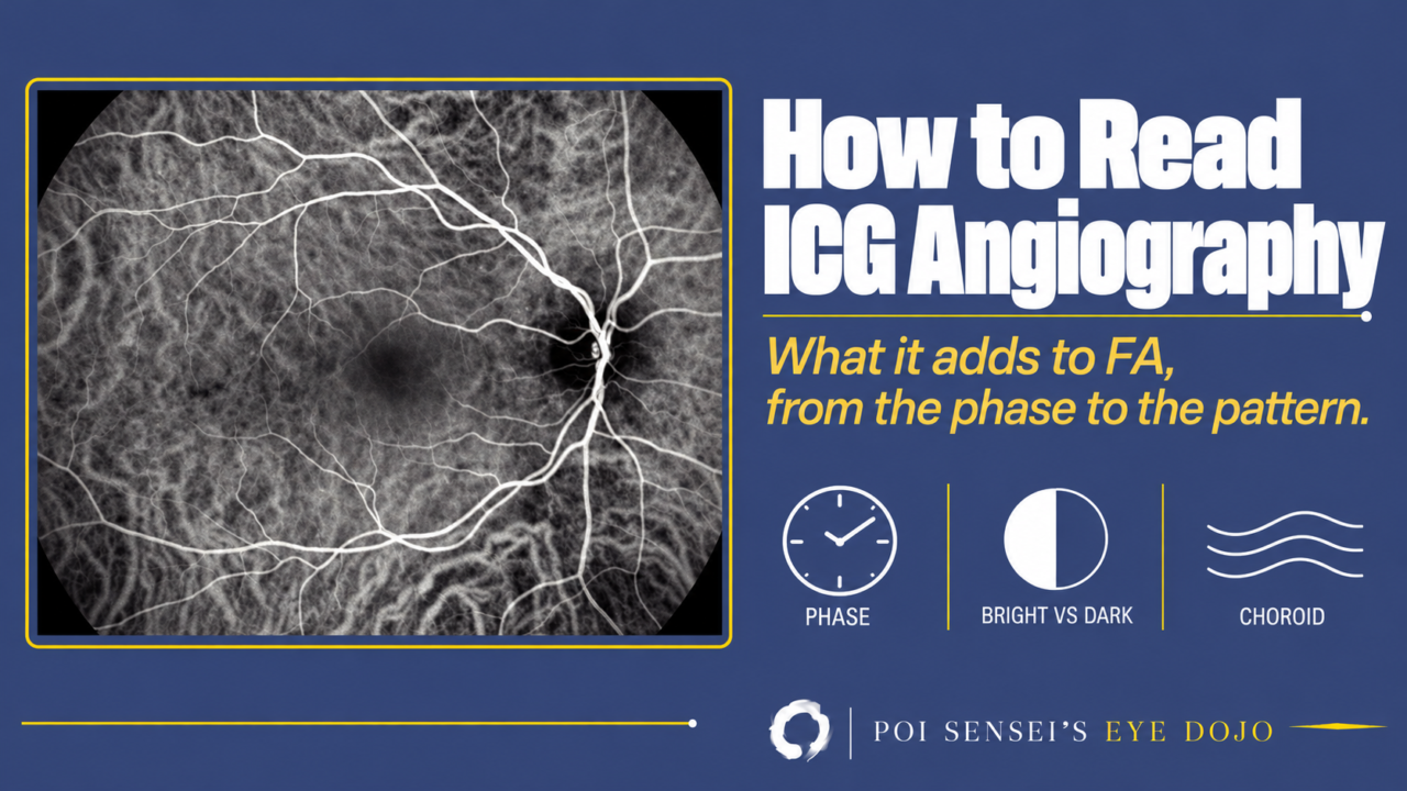
Fluorescein angiography (FA) shows the retinal vessels. Indocyanine green angiography (ICGA) shows the choroid behind them. Read it with the same first question as FA: is this area too bright or too dark? Then ask which choroidal disease gives that pattern.
- Know the phase: early, mid or late.
- Compare each area with the normal look for that phase: is it too bright or too dark?
- Match the pattern to a choroidal disease.
Why ICGA shows the choroid
On FA, the choroid is only a dim background glow. Two properties of indocyanine green change that.
- Almost all of the dye binds to proteins in the blood, so it stays in normal vessels. It hardly leaks out of the choriocapillaris.
- It glows in near-infrared light, while fluorescein glows in visible light. Near-infrared light passes through the retinal pigment epithelium (RPE), which acts like frosted glass on FA. It also passes through pigment and thin layers of blood.
So on ICGA you can see the choroidal vessels one by one. To see disease of the choroid, use ICGA.
When to add ICGA to FA
Add ICGA in these four situations.
- The choroid may be the problem: polypoidal choroidal vasculopathy (PCV), central serous chorioretinopathy (CSC) and other pachychoroid diseases, or uveitis that may involve the choroid.
- Blood, pigment, exudate or a cataract hides the view on FA. Near-infrared light passes through them and scatters less. Under a hemorrhage, video ICGA can even show a retinal arterial macroaneurysm pulsing.
- You need to see breaks in Bruch membrane, such as angioid streaks or lacquer cracks.
- FA cannot be used, for example after a past reaction to fluorescein.
ICGA does not replace FA for the retina. Leakage from the retinal capillaries and areas of closed capillaries hardly show on ICGA. For diabetic retinopathy and retinal vein occlusion, use FA.
Step 1: Know the phase
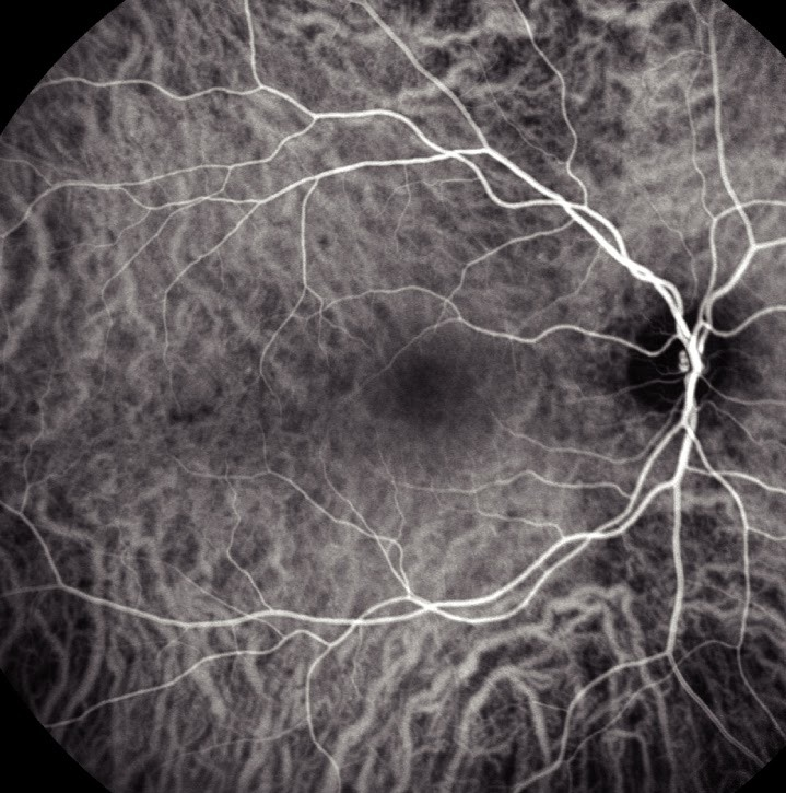
Early phase (the first minute). The choroidal arteries fill first, then the choroidal veins. If one area fills more than about 4 seconds later than the rest, think of poor choroidal flow. But even a healthy eye has watershed zones that fill a little late: a vertical band through the optic disc and a horizontal band through the disc and the fovea. Do not mistake them for disease. The small retinal vessels are hard to see, because the choroid behind them is so bright. Abnormal vessels, such as new vessels, start to show now.
Mid phase (about 3–15 minutes). A diffuse glow covers the macula and hides the vessels behind it. Outside the macula, dilated or tortuous choroidal veins and leakage stand out.
Late phase (up to about 45 minutes). The RPE has taken up some dye and glows evenly, so the vessels now look darker than the background. Anything still bright now is leakage or staining. Staining makes angioid streaks easier to see than on FA. In retinal vasculitis, inflamed vessel walls can stain now too (see the uveitis section below).
Late images matter more on ICGA than on FA.
Step 2: Too bright or too dark?
Compare each area with the same phase in a normal eye.
Too bright: when does it appear, and does it grow?
Not every bright area is disease. Where large choroidal vessels cross, the overlap is normally bright. For a truly bright area, ask when it appears and how it changes.
Abnormal vessels, such as polyps and new vessels, show from the early phase.
Leakage spreads with time. Leakage from the choroidal vessels (choroidal hyperpermeability) appears from the mid phase as a hazy bright patch. It often shows where FA has no leak at all.
A window defect is RPE atrophy that lets more light through. It is easy to miss in the early and mid phases, when the large choroidal vessels are bright and uneven. It gets clearer in the late phase and keeps the same size.
Staining means the tissue holds the dye. It stands out in the late phase as the background fades. Damaged vessel walls and scars stain.
Too dark: blocked, or not filling?
Too dark means one of two things: something in front is blocking the light (pigment, blood, fibrosis), or the dye is not reaching the area (poor flow). To tell them apart, look inside the dark area. If you can still see deeper choroidal vessels there, the flow is poor. If you see nothing at all, the light is blocked.
A block keeps the same shape and size in every phase. Early on, near-infrared light passes through thin blood or fluid, so a thin layer may not look dark at all. In the late phase, when the dye is weak, even the macular pigment or a serous retinal detachment can block it.
A filling defect is an area the dye does not reach. In uveitis it is usually many small dark spots that fade as the inflammation settles. In atrophy, such as a dystrophy or an old neovascular scar, the dark area stays sharp to the end. On FA the same scar turns bright, because fluorescein seeps in from around it. ICG does not.
One trap: a pigment epithelial detachment (PED) is bright on FA, but on ICGA it is mostly dark, with a bright rim. In age-related macular degeneration (AMD), look at a serous PED closely for hidden new vessels (see below).
Step 3: Match the pattern to the disease
Polypoidal choroidal vasculopathy: bright nodules
In PCV, the early phase shows bright nodules (polyps) at the ends of a branching vascular network of choroidal vessels. The network is a type 1 macular neovascularization (MNV) under the RPE. On FA the lesion is just a bright area. ICGA shows the polyps far better, so it is the key test for PCV.
Neovascular AMD: hot spot or plaque
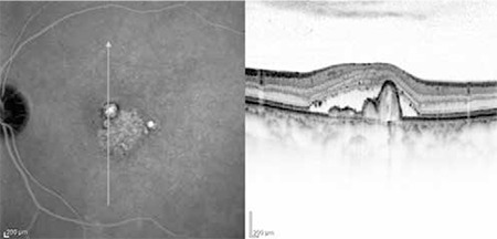
In neovascular AMD, FA remains the main angiogram, and ICGA is an add-on. It helps most when you suspect PCV, or when blood, fluid or pigment hides the MNV on FA. ICGA can still show the MNV as a small bright hot spot or a wider bright plaque. In a serous PED, this is how hidden MNV is often found. In type 3 MNV (retinal angiomatous proliferation), a hot spot in the mid or late phase usually makes the diagnosis.
VKH disease: dark dots
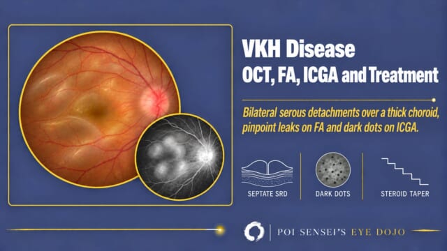
In acute Vogt-Koyanagi-Harada (VKH) disease, the inflamed choroid shows scattered dark dots. The large choroidal vessels look fuzzy, and the optic disc is bright. The dark dots fade with treatment and come back when the disease recurs, so ICGA also helps you follow the disease.
Pachychoroid and CSC: patchy bright areas
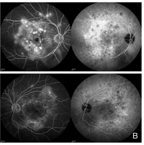
In CSC and other pachychoroid diseases (a thick choroid with dilated, leaky vessels), the early phase shows dilated choroidal veins. Call a vein dilated if it is wider near the macula than farther out, if its width swells and narrows like a string of sausages, or if it is wider than the same vein in the other eye. On wide-field ICGA, the dilated veins can often be followed all the way to congested vortex veins.
In the mid and late phases, patchy bright areas with blurred edges appear where the choroidal vessels are leaky (choroidal hyperpermeability). They often appear where FA shows no leak at all, and in places that cause no symptoms.
ICGA helps most in chronic CSC, where the leak on FA is often hard to read. ICGA still shows the leaky choroid and the dilated veins. The bright areas also guide photodynamic therapy (PDT), and eyes that show them tend to respond well.
Uveitis: what ICGA adds
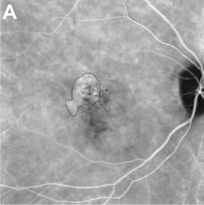
In uveitis, FA shows the retina and its vessels, and ICGA shows the choroid. Take both when the choroid may be inflamed. ICGA helps in three ways.
Two patterns of choroiditis
ICGA tells you which layer of the choroid is inflamed.
Choriocapillaritis (inflammation of the choriocapillaris) gives dark patches of different sizes. They are dark from the early phase and clearest in the late phase. There are often more of them than you can see on the fundus. Examples are acute posterior multifocal placoid pigment epitheliopathy (APMPPE), serpiginous choroiditis and multiple evanescent white dot syndrome (MEWDS). See also white dot syndromes.
Stromal choroiditis (inflammation of the choroidal stroma) gives small, round, evenly spread dark dots from the early phase. The large choroidal vessels look fuzzy and leak, so the late phase shows a diffuse glow. VKH disease and sympathetic ophthalmia start in the choroid. Sarcoidosis, tuberculosis and syphilis can inflame it too. In birdshot chorioretinopathy, the dark dots far outnumber the spots on the fundus and do not match them.
So sharp, patchy dark areas point to the choriocapillaris. Faint dots spread evenly over the whole fundus, with fuzzy large vessels, point to the stroma.
Follow-up: ICGA clears last
ICGA lesions often take longer to clear than the signs on the fundus and FA. In APMPPE, if you stop treatment while dark patches remain on ICGA, the disease often comes back. When it recurs, new dark patches can appear on ICGA before symptoms, and before any change on the fundus or FA. In VKH disease too, ICGA can show leakage in the choroid and dark dots for months after the eye looks quiet. So follow choroiditis with ICGA, not FA alone.
Retinal vasculitis: check the late phase
FA remains the main test for retinal vasculitis. Fluorescein leaks out of even mildly inflamed vessels, and FA also shows closed capillaries. ICGA shows neither well.
ICGA adds its late phase. By then the dye has left the retinal vessels, so an inflamed vessel wall that has taken up dye can stand out as a bright line.
Which vessels are inflamed matters. Most retinal vasculitis affects the veins (periphlebitis), as in sarcoidosis, tuberculosis and multiple sclerosis. Arteritis is less common. When the arteries are inflamed, think of infection first, such as acute retinal necrosis (ARN) or toxoplasmosis. Behçet disease and lupus can also inflame the arteries.
ICGA also shows whether the choroid is inflamed, as can happen in sarcoidosis and tuberculosis.
Before you inject
If the patient has reacted to indocyanine green before, do not give it. The dye contains a small amount of iodide, so also ask about iodine allergy.
ICGA is generally better tolerated than FA. Nausea and hives are uncommon, and serious reactions are very rare, but anaphylaxis probably occurs about as often as with FA. The dye is cleared by the liver, so take care in liver disease. Its safety in pregnancy has not been established.
Summary
- ICGA shows the choroid, but FA is still the test for the retinal capillaries.
- Know the phase, then compare each area with the normal look for that phase.
- Note when a bright area appears and whether it grows, and whether deeper vessels show inside a dark one.
- Bright nodules suggest PCV, dark dots or patches suggest choroiditis, and patchy bright areas suggest pachychoroid.
- In uveitis, ICGA lesions often outlast the signs on FA, so follow the choroid with ICGA.

Which retinal layer makes which wave, the three abnormal patterns, and the dark-adaptation trap to check before you report “negative” — on one page, by email. One email when I publish something new, about once a month. Unsubscribe anytime.