Autosomal Recessive Bestrophinopathy (ARB): Fundus, OCT and EOG
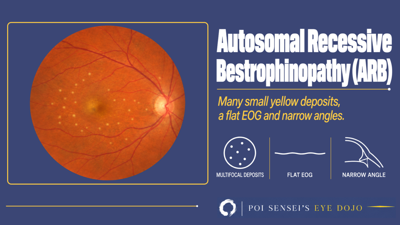
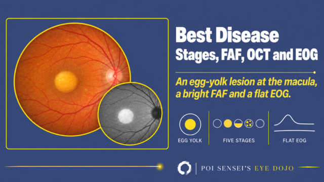
Both eyes show multiple small yellow deposits scattered over the posterior pole. OCT shows shallow subretinal fluid with cystoid spaces in the retina, and the EOG shows almost no light rise. Think of autosomal recessive bestrophinopathy (ARB). This article shows how ARB looks on the fundus, autofluorescence and OCT, how the EOG and ERG separate it from Best disease, and why you must check the angle.
- Vision falls between childhood and middle age.
- Multiple small yellow deposits sit around the macula and the optic disc in both eyes.
- OCT shows shallow subretinal fluid and cystoid spaces in the retina.
- The EOG is severely reduced, and the full-field ERG is often abnormal too.
- The eyes are hyperopic, with shallow anterior chambers.
What is ARB?
ARB is an autosomal recessive retinal dystrophy caused by mutations in both copies of the BEST1 gene. This is the same gene as in Best disease. The inheritance and the fundus are different, so ARB is regarded as a separate disease. In both diseases, the retinal pigment epithelium (RPE) does not work properly, and lipofuscin builds up.
Best disease is autosomal dominant, so one parent is often affected. In ARB, the parents are carriers, and their fundus and EOG are normal[1].
Symptoms
Typical patients come in with reduced vision at about 10 to 40 years of age, though it can start in early childhood or after 60. Children often still have good central vision. Vision then falls slowly over the years[2].
Findings
Fundus and autofluorescence
ARB does not usually show the single egg-yolk (vitelliform) lesion of Best disease. Instead, many small, round, yellow deposits are scattered over the posterior pole, often around the optic disc as well as the macula. They come from lipofuscin, so they are bright (hyperautofluorescent) on fundus autofluorescence.
Over time, some eyes develop atrophy or fibrosis at the macula[2].
OCT
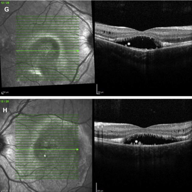
OCT shows shallow subretinal fluid (a serous retinal detachment) and cystoid spaces within the retina that can look like schisis. The yellow deposits seen on the fundus sit under the retina as bright material. The subretinal fluid usually stays for years[2].
EOG
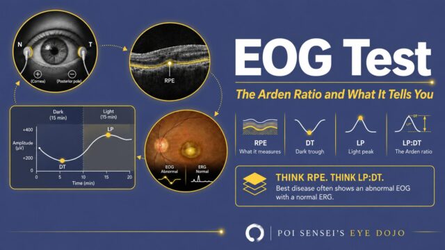
As in Best disease, the RPE is abnormal, so the electro-oculogram (EOG) is reduced. In ARB, the light rise is severely reduced or absent at every age[2]. Rarely, it is close to normal[1].
ERG
In Best disease, the full-field electroretinogram (ERG) is normal. In ARB, it is often abnormal. It can still be normal in children, so a normal ERG does not rule out ARB[2].
Complications
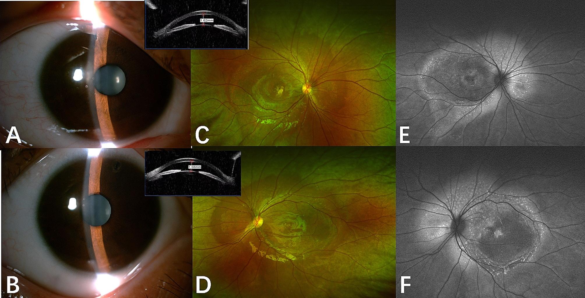
ARB eyes are often hyperopic, with a short axial length, a shallow anterior chamber and a narrow angle. Angle-closure glaucoma is common. Some patients are first diagnosed with glaucoma, and the retinal disease is found later[1].
Summary
- ARB is caused by mutations in both copies of BEST1, the gene of Best disease.
- Look for multiple small yellow deposits that are bright on autofluorescence, and shallow subretinal fluid with cystoid spaces on OCT.
- The EOG is severely reduced, and unlike in Best disease, the full-field ERG is often abnormal.
- The eyes are often hyperopic with narrow angles, so check the angle.
References
[1] Zhao D, Gu VY, Wang Y, Peng J, Lyu J, Fei P, Xu Y, Zhang X, Zhao P. Clinical and genetic features in autosomal recessive bestrophinopathy in Chinese cohort. BMC Ophthalmol. 2024;24(1):308. PMID 39048936.
[2] Casalino G, Khan KN, Armengol M, Wright G, Pontikos N, Georgiou M, Webster AR, Robson AG, Grewal PS, Michaelides M. Autosomal recessive bestrophinopathy: clinical features, natural history, and genetic findings in preparation for clinical trials. Ophthalmology. 2021;128(5):706-718. PMID 33039401.
Which retinal layer makes which wave, the three abnormal patterns, and the dark-adaptation trap to check before you report “negative” — on one page, by email. One email when I publish something new, about once a month. Unsubscribe anytime.