Acquired Vitelliform Lesion (AVL): OCT, Autofluorescence and How Not to Mistake It for Wet AMD
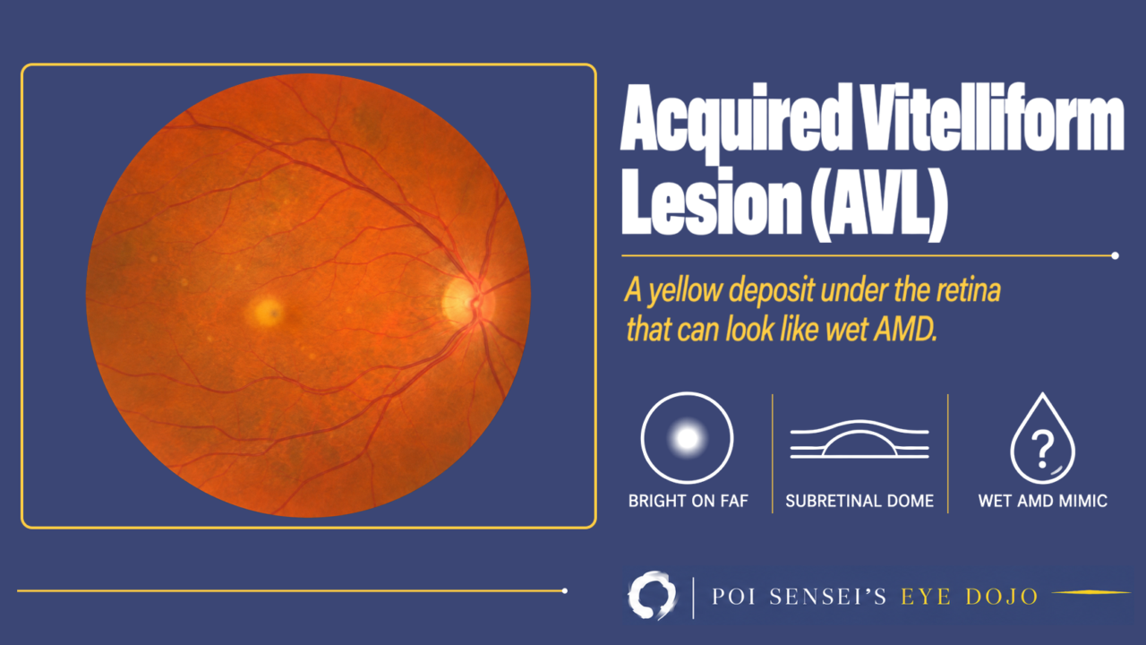
An older patient has a small yellow deposit at the macula. OCT shows a dome of bright material under the retina, sometimes with a little fluid next to it. Before you call it wet age-related macular degeneration (AMD), think of an acquired vitelliform lesion (AVL). This article explains what the yellow material is, what causes it, how it looks on autofluorescence, OCT and angiography, and how to tell it from wet AMD and Best disease.
- An older adult has a yellow deposit at the macula, often with drusen.
- The deposit is bright on fundus autofluorescence.
- OCT shows bright material between the retina and the retinal pigment epithelium (RPE).
- There is no sign of new vessels, even when a little fluid is present.
What is an AVL?
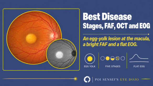
Vitelliform means "like an egg yolk." A vitelliform lesion is yellow material that collects in the subretinal space, between the photoreceptors and the RPE.
In a child or young adult, this picture usually means Best disease, an inherited macular dystrophy. An acquired vitelliform lesion appears in adult life as a result of another macular disease.
Why the material builds up
Photoreceptors shed the tips of their outer segments every day, and the RPE takes them up and digests them. To do this, the RPE must be in contact with the tips. When the retina is separated from the RPE, the shed material is not cleared. The same happens when the RPE is not working well. The material then piles up under the retina[1].
Under the microscope, the deposit is a mix of outer segment debris, RPE pigment granules, lipid and other material[2]. On the fundus, this material looks yellow.
Causes
Many diseases can cause an AVL. What they share is poor contact between the photoreceptors and the RPE, or an RPE that is not working well[3].
- AMD with large drusen or a pigment epithelial detachment
- Cuticular drusen and subretinal drusenoid deposits
- Vitreomacular traction and epiretinal membrane
- Central serous chorioretinopathy (CSC)
Findings
Fundus
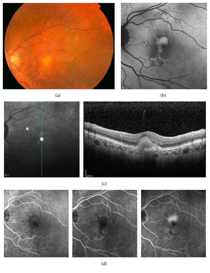
A slightly raised yellow deposit lies under or near the fovea. It is round or irregular and often about a third of a disc diameter across. A small pigment spot may sit in the middle[2]. Drusen are often seen around it. One or both eyes can be affected, and the lesion may sit off the center of the fovea.
Fundus autofluorescence
The deposit is intensely bright (hyperautofluorescent)[1]. When the lesion later turns into atrophy, the area becomes dark.
OCT
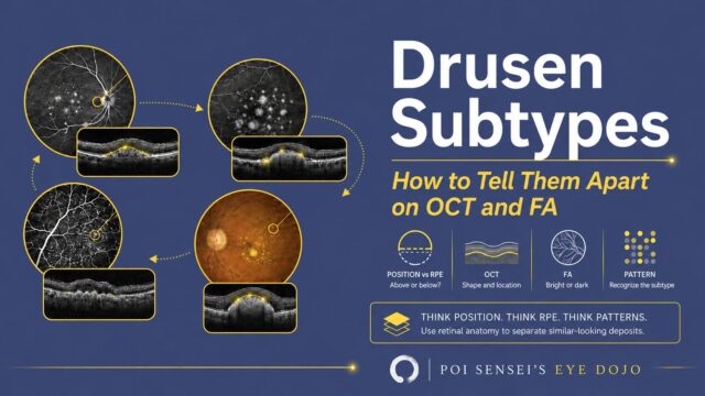
OCT is the key test. The yellow material is a dome of bright (hyperreflective) material in the subretinal space, above the RPE and below the photoreceptors. This location separates it from ordinary drusen and pigment epithelial detachments, which lie under the RPE.
A dark (hyporeflective) space sometimes sits next to the material[1]. It may be fluid, but it does not always mean active neovascularization[2].
Vision depends on the photoreceptors over the deposit. If the external limiting membrane and the ellipsoid zone under the fovea are intact, vision tends to be good[1].
Fluorescein angiography
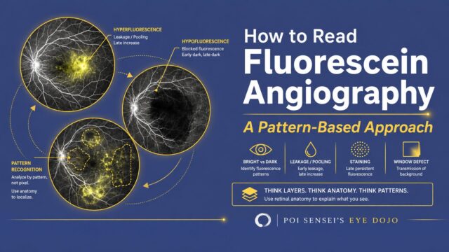
On fluorescein angiography, the deposit blocks the fluorescence in the early phase and looks dark. In the late phase, it stains and looks bright. This late brightness can look like leakage from macular neovascularization (MNV), abnormal new vessels under the retina, formerly called choroidal neovascularization (CNV)[1]. Even true leakage can appear without MNV, so late brightness alone does not prove MNV[5].
OCT angiography
OCT angiography shows blood flow without dye. It helps check for new vessels under the lesion[2].
How the lesion changes over time
An AVL can pass through stages like those of Best disease: vitelliform (egg yolk), pseudohypopyon (the material settles with a flat upper edge), vitelliruptive (scrambled egg) and atrophic. Not every lesion goes through all of them[3].
The lesion grows, reaches a peak and then shrinks. In a study of AVLs in AMD, about half of them collapsed into atrophy of the RPE and outer retina. The rest stayed or disappeared without atrophy[2].
New vessels can also develop. In one large series, about 1 in 8 eyes with AVL in AMD developed MNV, mostly after the lesion had started to shrink[4].
Telling AVL from other diseases
Wet AMD
Yellow material, a little subretinal fluid on OCT and late brightness on angiography can look just like wet AMD. Anti-VEGF injections treat MNV. But in AVL without MNV, the fluid often comes and goes on its own, and the injections do not clearly help[5].
Best disease and adult-onset vitelliform dystrophy
Adult-onset foveomacular vitelliform dystrophy (AOFVD) is a macular dystrophy that also forms a yellow deposit at the macula, usually after age 40. Some patients have no family history. It looks much like AVL.
On the fundus alone, Best disease, AOFVD and AVL are hard to tell apart. Age, the other eye, other macular disease and the electrooculogram (EOG), which tests RPE function, help[6].
| Best | AOFVD | AVL | |
|---|---|---|---|
| Onset | Childhood | After 40 | Older age |
| Eyes | Usually both | Usually both | One or both |
| Lesion | Central | Central, round | Not always central |
| Other disease | None | None | Drusen, traction, CSC |
| EOG | Abnormal | Normal or mildly low | Normal |
Treatment
No treatment removes the yellow material. Follow the lesion with OCT and autofluorescence. If MNV develops, it is treated with anti-VEGF injections.
Summary
- An AVL is yellow material under the retina that appears in adults with another macular disease, such as AMD.
- It is bright on autofluorescence and sits above the RPE on OCT.
- Fluid and late staining can look like wet AMD. Look for MNV before injecting.
- No treatment removes the material. Anti-VEGF injections are for MNV.
References
[1] Freund KB, Laud K, Lima LH, Spaide RF, Zweifel S, Yannuzzi LA. Acquired vitelliform lesions: correlation of clinical findings and multiple imaging analyses. Retina. 2011;31(1):13-25. PMID 21102371.
[2] Brinkmann M, Bacci T, Kar D, Messinger JD, Sloan KR, Chen L, Hamann T, Wiest M, Freund KB, Zweifel S, Curcio CA. Histology and clinical lifecycle of acquired vitelliform lesion, a pathway to advanced age-related macular degeneration. Am J Ophthalmol. 2022;240:99-114. PMID 35192790.
[3] Rocha Bastos R, Ferreira CS, Brandão E, Falcão-Reis F, Carneiro ÂM. Multimodal image analysis in acquired vitelliform lesions and adult-onset foveomacular vitelliform dystrophy. J Ophthalmol. 2016;2016:6037537. PMID 27190637.
[4] Balaratnasingam C, Hoang QV, Inoue M, Curcio CA, Dolz-Marco R, Yannuzzi NA, Dhrami-Gavazi E, Yannuzzi LA, Freund KB. Clinical characteristics, choroidal neovascularization, and predictors of visual outcomes in acquired vitelliform lesions. Am J Ophthalmol. 2016;172:28-38. PMID 27640006.
[5] Juliano J, Patel S, Ameri H. Acquired vitelliform macular degeneration: characteristics and challenges of managing subretinal fluid. J Ophthalmic Vis Res. 2021;16(4):582-591. PMID 34840681.
[6] Salmon JF. Kanski’s Clinical Ophthalmology: A Systematic Approach. 10th ed. Elsevier; 2025. Chapter 15, Inherited retinal degenerations: Adult-onset vitelliform macular dystrophy.
Which retinal layer makes which wave, the three abnormal patterns, and the dark-adaptation trap to check before you report “negative” — on one page, by email. One email when I publish something new, about once a month. Unsubscribe anytime.